 Movie
Movie Controller

[English] 日本語
 Yorodumi
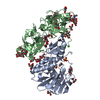
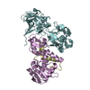
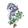
- PDB-5y97: Crystal structure of snake gourd seed lectin in complex with lactose -
+ Open data

- Basic information
| Entry | Database: PDB / ID: 5y97 | |||||||||
|---|---|---|---|---|---|---|---|---|---|---|
| Title | Crystal structure of snake gourd seed lectin in complex with lactose | |||||||||
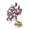
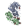
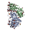
 Components Components | (Seed lectin) x 3 | |||||||||
 Keywords Keywords | PLANT PROTEIN / beta-trefoil / Lectin | |||||||||
| Function / homology |  Function and homology information Function and homology informationrRNA N-glycosylase activity / carbohydrate binding / negative regulation of translation Similarity search - Function | |||||||||
| Biological species |  Trichosanthes anguina (plant) Trichosanthes anguina (plant) | |||||||||
| Method |  X-RAY DIFFRACTION / SYNCHROTRON / MOLECULAR REPLACEMENT / Resolution: 3.05 Å X-RAY DIFFRACTION / SYNCHROTRON / MOLECULAR REPLACEMENT / Resolution: 3.05 Å | |||||||||
 Authors Authors | Chandran, T. / Vijayan, M. / Sivaji, N. / Surolia, A. | |||||||||
| Funding support |  India, 1items India, 1items
| |||||||||
 Citation Citation | Journal: Glycobiology / Year: 2018 Title: Ligand binding and retention in snake gourd seed lectin (SGSL). A crystallographic, thermodynamic and molecular dynamics study Authors: Chandran, T. / Sivaji, N. / Surolia, A. / Vijayan, M. #1: Journal: Acta Crystallogr. D Biol. Crystallogr. / Year: 2013Title: The sequence and structure of snake gourd (Trichosanthes anguina) seed lectin, a three-chain nontoxic homologue of type II RIPs. Authors: Sharma, A. / Pohlentz, G. / Bobbili, K.B. / Jeyaprakash, A.A. / Chandran, T. / Mormann, M. / Swamy, M.J. / Vijayan, M. | |||||||||
| History |
|
- Structure visualization
| Structure viewer | Molecule: MolmilJmol/JSmol |
|---|
- Downloads & links
-Download
| PDBx/mmCIF format | 5y97.cif.gz | 221.9 KB | Display | PDBx/mmCIF format |
|---|---|---|---|---|
| PDB format | pdb5y97.ent.gz | 179.8 KB | Display | PDB format |
| PDBx/mmJSON format | 5y97.json.gz | Tree view | PDBx/mmJSON format | |
| Others |  Other downloads Other downloads |
-Validation report
| Arichive directory | https://data.pdbj.org/pub/pdb/validation_reports/y9/5y97ftp://data.pdbj.org/pub/pdb/validation_reports/y9/5y97 | HTTPS FTP |
|---|
-Related structure data
| Related structure data | 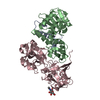 5y42C  4hr6S S: Starting model for refinement C: citing same article ( |
|---|---|
| Similar structure data |


-Links
 PDBj
PDBj


- Assembly
| Deposited unit | 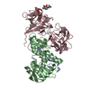
| ||||||||
|---|---|---|---|---|---|---|---|---|---|
| 1 |
| ||||||||
| Unit cell |
|
-Components
-Protein , 2 types, 2 molecules BC
| #2: Protein | Mass: 23387.219 Da / Num. of mol.: 1 / Fragment: UNP residues 47-255 / Source method: isolated from a natural source / Source: (natural) Trichosanthes anguina (plant) / References: UniProt: U3KRF8 |
|---|---|
| #3: Protein | Mass: 29287.674 Da / Num. of mol.: 1 / Fragment: UNP residues 256-519 / Source method: isolated from a natural source / Source: (natural) Trichosanthes anguina (plant) / References: UniProt: U3KRF8 |
-Protein/peptide / Non-polymers , 2 types, 6 molecules A
| #1: Protein/peptide | Mass: 4647.276 Da / Num. of mol.: 1 / Fragment: UNP residues 4-44 / Source method: isolated from a natural source / Source: (natural) Trichosanthes anguina (plant) / References: UniProt: U3KRF8 |
|---|---|
| #6: Water | ChemComp-HOH / Mass: 18.015 Da / Num. of mol.: 5 / Source method: isolated from a natural source / Formula: H2O |
-Sugars , 2 types, 3 molecules 
| #4: Polysaccharide |   Source method: isolated from a genetically manipulated source Details: oligosaccharide / References: beta-lactose #5: Sugar | ChemComp-NAG / |  Type: D-saccharide, beta linking / Mass: 221.208 Da / Num. of mol.: 1 Type: D-saccharide, beta linking / Mass: 221.208 Da / Num. of mol.: 1Source method: isolated from a genetically manipulated source Formula: C8H15NO6 |
|---|
-Details
| Has protein modification | Y |
|---|---|
| Sequence details | There are three different polypeptide chains. A and B are contiguous and are results from cleavage. ...There are three different polypeptide chains. A and B are contiguous and are results from cleavage. C is an independent chain. |
-Experimental details
-Experiment
| Experiment | Method: X-RAY DIFFRACTION / Number of used crystals: 1 |
|---|
- Sample preparation
| Crystal | Density Matthews: 3.51 Å3/Da / Density % sol: 64.93 % / Description: hexagonal crystals |
|---|---|
| Crystal grow | Temperature: 300 K / Method: vapor diffusion, hanging drop / pH: 7.8 / Details: reservoir solution contained 4M sodium formate / PH range: 6.4-7.9 |
-Data collection
| Diffraction | Mean temperature: 100 K / Ambient temp details: liquid nitrogen |
|---|---|
| Diffraction source | Source: SYNCHROTRON / Site: ESRF  / Beamline: BM14 / Wavelength: 0.9537 Å / Beamline: BM14 / Wavelength: 0.9537 Å |
| Detector | Type: MARMOSAIC 225 mm CCD / Detector: CCD / Date: Jul 14, 2015 |
| Radiation | Monochromator: si (111) / Protocol: SINGLE WAVELENGTH / Monochromatic (M) / Laue (L): M / Scattering type: x-ray |
| Radiation wavelength | Wavelength: 0.9537 Å / Relative weight: 1 |
| Reflection | Resolution: 3.05→94.77 Å / Num. obs: 16493 / % possible obs: 100 % / Redundancy: 18.9 % / CC1/2: 0.99 / Rmerge(I) obs: 0.146 / Net I/σ(I): 13.3 |
| Reflection shell | Resolution: 3.05→3.21 Å / Redundancy: 19.5 % / Rmerge(I) obs: 1.534 / Mean I/σ(I) obs: 2 / Num. unique obs: 2340 / CC1/2: 0.808 / % possible all: 100 |
- Processing
| Software |
| ||||||||||||||||||||||||||||||||||||||||||||||||||||||||||||||||||||||||||||||||||||||||||||||||||||||||||||||||||||||||||||||||||||||||||||||||||||||||||||||||||||||||||||||||||||||
|---|---|---|---|---|---|---|---|---|---|---|---|---|---|---|---|---|---|---|---|---|---|---|---|---|---|---|---|---|---|---|---|---|---|---|---|---|---|---|---|---|---|---|---|---|---|---|---|---|---|---|---|---|---|---|---|---|---|---|---|---|---|---|---|---|---|---|---|---|---|---|---|---|---|---|---|---|---|---|---|---|---|---|---|---|---|---|---|---|---|---|---|---|---|---|---|---|---|---|---|---|---|---|---|---|---|---|---|---|---|---|---|---|---|---|---|---|---|---|---|---|---|---|---|---|---|---|---|---|---|---|---|---|---|---|---|---|---|---|---|---|---|---|---|---|---|---|---|---|---|---|---|---|---|---|---|---|---|---|---|---|---|---|---|---|---|---|---|---|---|---|---|---|---|---|---|---|---|---|---|---|---|---|---|
| Refinement | Method to determine structure: MOLECULAR REPLACEMENT Starting model: 4HR6 Resolution: 3.05→94.77 Å / Cor.coef. Fo:Fc: 0.938 / Cor.coef. Fo:Fc free: 0.909 / SU B: 45.997 / SU ML: 0.34 / Cross valid method: THROUGHOUT / ESU R Free: 0.395 / Details: HYDROGENS HAVE BEEN ADDED IN THE RIDING POSITIONS
| ||||||||||||||||||||||||||||||||||||||||||||||||||||||||||||||||||||||||||||||||||||||||||||||||||||||||||||||||||||||||||||||||||||||||||||||||||||||||||||||||||||||||||||||||||||||
| Solvent computation | Ion probe radii: 0.8 Å / Shrinkage radii: 0.8 Å / VDW probe radii: 1.2 Å | ||||||||||||||||||||||||||||||||||||||||||||||||||||||||||||||||||||||||||||||||||||||||||||||||||||||||||||||||||||||||||||||||||||||||||||||||||||||||||||||||||||||||||||||||||||||
| Displacement parameters | Biso mean: 118.829 Å2
| ||||||||||||||||||||||||||||||||||||||||||||||||||||||||||||||||||||||||||||||||||||||||||||||||||||||||||||||||||||||||||||||||||||||||||||||||||||||||||||||||||||||||||||||||||||||
| Refinement step | Cycle: 1 / Resolution: 3.05→94.77 Å
| ||||||||||||||||||||||||||||||||||||||||||||||||||||||||||||||||||||||||||||||||||||||||||||||||||||||||||||||||||||||||||||||||||||||||||||||||||||||||||||||||||||||||||||||||||||||
| Refine LS restraints |
|