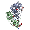
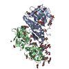
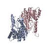
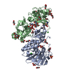
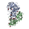
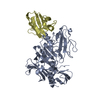
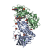
登録情報 データベース : PDB / ID : 4hr6タイトル Crystal structure of snake gourd (Trichosanthes anguina) seed lectin, a three chain homologue of type II RIPs (LECTIN) x 3 キーワード / / / / / 機能・相同性 分子機能 ドメイン・相同性 構成要素
/ / / / / / / / / / / / / / / / / / / / / / / / / / / / / / / / / / / / / / / 生物種 Trichosanthes anguina (植物)手法 / / / 解像度 : 2.25 Å データ登録者 Sharma, A. / Pohlentz, G. / Bobbili, K.B. / Jeyaprakash, A.A. / Chandran, T. / Mormann, M. / Swamy, M.J. / Vijayan, M. ジャーナル : Acta Crystallogr.,Sect.D / 年 : 2013タイトル : The sequence and structure of snake gourd (Trichosanthes anguina) seed lectin, a three-chain nontoxic homologue of type II RIPs.
著者 :
Sharma, A. / Pohlentz, G. / Bobbili, K.B. / Jeyaprakash, A.A. / Chandran, T. / Mormann, M. / Swamy, M.J. / Vijayan, M. 履歴 登録 2012年10月26日 登録サイト / 処理サイト 改定 1.0 2013年8月7日 Provider / タイプ 改定 1.1 2019年12月25日 Group / カテゴリ Item _citation.country / _citation.journal_id_CSD ... _citation.country / _citation.journal_id_CSD / _citation.journal_id_ISSN / _citation.pdbx_database_id_PubMed / _citation.title 改定 1.2 2020年7月29日 Group / Derived calculations / Structure summaryカテゴリ chem_comp / entity ... chem_comp / entity / pdbx_chem_comp_identifier / pdbx_entity_nonpoly / struct_site / struct_site_gen Item _chem_comp.mon_nstd_flag / _chem_comp.name ... _chem_comp.mon_nstd_flag / _chem_comp.name / _chem_comp.type / _entity.pdbx_description / _pdbx_entity_nonpoly.name 解説 / Provider / タイプ 改定 1.3 2024年11月20日 Group / Database references / Structure summaryカテゴリ chem_comp / chem_comp_atom ... chem_comp / chem_comp_atom / chem_comp_bond / database_2 / pdbx_entry_details / pdbx_modification_feature Item _chem_comp.pdbx_synonyms / _database_2.pdbx_DOI ... _chem_comp.pdbx_synonyms / _database_2.pdbx_DOI / _database_2.pdbx_database_accession / _pdbx_entry_details.has_protein_modification
すべて表示 表示を減らす
 ムービー
ムービー コントローラー


 データを開く
 基本情報
基本情報 要素
要素 キーワード
キーワード 機能・相同性情報
 Trichosanthes anguina (植物)
 X線回折 /
  データ登録者
 引用
引用 構造の表示
構造の表示 ダウンロードとリンク
 その他のダウンロード
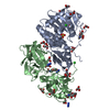


 PDBj
PDBj

 集合体
集合体

 タイプ: D-saccharide / 分子量: 194.182 Da / 分子数: 2 / 由来タイプ: 組換発現 / 式: C7H14O6
 分子量: 18.015 Da / 分子数: 178 / 由来タイプ: 天然 / 式: H2O
 試料調製
試料調製 / ビームライン: 5.2R / 波長: 1 Å
 解析
解析