 Movie
Movie Controller

[English] 日本語
 Yorodumi
- PDB-2r9k: Crystal Structure of Misteltoe Lectin I in Complex with Phloretamide -
+ Open data

- Basic information
| Entry | Database: PDB / ID: 2r9k | ||||||||||||
|---|---|---|---|---|---|---|---|---|---|---|---|---|---|
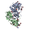
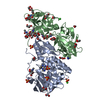
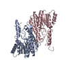
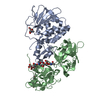
| Title | Crystal Structure of Misteltoe Lectin I in Complex with Phloretamide | ||||||||||||
 Components Components | (Beta-galactoside-specific lectin ...) x 2 | ||||||||||||
 Keywords Keywords | HYDROLASE / ML-I / phloretamide / Viscum album / Glycoprotein / Lectin / Plant defense / Protein synthesis inhibitor / Toxin | ||||||||||||
| Function / homology |  Function and homology information Function and homology informationrRNA N-glycosylase / rRNA N-glycosylase activity / defense response / toxin activity / carbohydrate binding / negative regulation of translation Similarity search - Function | ||||||||||||
| Biological species |  Viscum album (European mistletoe) Viscum album (European mistletoe) | ||||||||||||
| Method |  X-RAY DIFFRACTION / SYNCHROTRON / MOLECULAR REPLACEMENT / Resolution: 2.7 Å X-RAY DIFFRACTION / SYNCHROTRON / MOLECULAR REPLACEMENT / Resolution: 2.7 Å | ||||||||||||
 Authors Authors | Meyer, A. / Rypniewski, W. / Celewicz, L. / Erdmann, V.A. / Voelter, W. / Betzel, C. | ||||||||||||
 Citation Citation | Journal: Biochem.Biophys.Res.Commun. / Year: 2007 Title: The mistletoe lectin I--phloretamide structure reveals a new function of plant lectins. Authors: Meyer, A. / Rypniewski, W. / Celewicz, L. / Erdmann, V.A. / Voelter, W. / Singh, T.P. / Genov, N. / Barciszewski, J. / Betzel, C.h. | ||||||||||||
| History |
| ||||||||||||
| Remark 999 | The authors stae that the plant proteins can differ in some codons depending on the season and on ...The authors stae that the plant proteins can differ in some codons depending on the season and on the host where the mistletoe has grown. |
- Structure visualization
| Structure viewer | Molecule: MolmilJmol/JSmol |
|---|
- Downloads & links
-Download
| PDBx/mmCIF format | 2r9k.cif.gz | 118.6 KB | Display | PDBx/mmCIF format |
|---|---|---|---|---|
| PDB format | pdb2r9k.ent.gz | 91 KB | Display | PDB format |
| PDBx/mmJSON format | 2r9k.json.gz | Tree view | PDBx/mmJSON format | |
| Others |  Other downloads Other downloads |
-Validation report
| Arichive directory | https://data.pdbj.org/pub/pdb/validation_reports/r9/2r9kftp://data.pdbj.org/pub/pdb/validation_reports/r9/2r9k | HTTPS FTP |
|---|
-Related structure data
| Related structure data |  1m2tS S: Starting model for refinement |
|---|---|
| Similar structure data |


-Links
 PDBj
PDBj

- Assembly
| Deposited unit | 
| ||||||||||||||||||||||||||||||||||||
|---|---|---|---|---|---|---|---|---|---|---|---|---|---|---|---|---|---|---|---|---|---|---|---|---|---|---|---|---|---|---|---|---|---|---|---|---|---|
| 1 |
| ||||||||||||||||||||||||||||||||||||
| 2 | 
| ||||||||||||||||||||||||||||||||||||
| 3 | 
| ||||||||||||||||||||||||||||||||||||
| Unit cell |
| ||||||||||||||||||||||||||||||||||||
| Components on special symmetry positions |
| ||||||||||||||||||||||||||||||||||||
| Noncrystallographic symmetry (NCS) | NCS domain:
NCS domain segments: Dom-ID: 1 / Component-ID: 1 / Refine code: 1
NCS ensembles :
|
-Components
-Beta-galactoside-specific lectin ... , 2 types, 2 molecules AB
| #1: Protein | Mass: 27820.066 Da / Num. of mol.: 1 Fragment: Beta-galactoside-specific lectin 1 chain A isoform 1, UNP residues 34-287 Source method: isolated from a genetically manipulated source Source: (gene. exp.) Viscum album (European mistletoe) / References: UniProt: P81446*PLUS |
|---|---|
| #2: Protein | Mass: 28568.939 Da / Num. of mol.: 1 Fragment: Beta-galactoside-specific lectin 1 chain B, UNP residues 302-564 Source method: isolated from a genetically manipulated source Source: (gene. exp.) Viscum album (European mistletoe) / References: UniProt: P81446 |
-Sugars , 2 types, 5 molecules 
| #3: Polysaccharide | 2-acetamido-2-deoxy-beta-D-glucopyranose-(4-4)-2-acetamido-2-deoxy-beta-D-glucopyranose Type: oligosaccharide / Mass: 424.401 Da / Num. of mol.: 1 Source method: isolated from a genetically manipulated source |
|---|---|
| #4: Sugar | ChemComp-NAG /  Type: D-saccharide, beta linking / Mass: 221.208 Da / Num. of mol.: 4 Type: D-saccharide, beta linking / Mass: 221.208 Da / Num. of mol.: 4Source method: isolated from a genetically manipulated source Formula: C8H15NO6 |
-Non-polymers , 5 types, 82 molecules 


| #5: Chemical | ChemComp-SO4 /  Mass: 96.063 Da / Num. of mol.: 4 / Source method: obtained synthetically / Formula: SO4 Mass: 96.063 Da / Num. of mol.: 4 / Source method: obtained synthetically / Formula: SO4#6: Chemical | ChemComp-GOL /  Mass: 92.094 Da / Num. of mol.: 4 / Source method: obtained synthetically / Formula: C3H8O3 Mass: 92.094 Da / Num. of mol.: 4 / Source method: obtained synthetically / Formula: C3H8O3#7: Chemical | ChemComp-CL / |  Mass: 35.453 Da / Num. of mol.: 1 / Source method: obtained synthetically / Formula: Cl Mass: 35.453 Da / Num. of mol.: 1 / Source method: obtained synthetically / Formula: Cl#8: Chemical | ChemComp-SGI / |  Mass: 165.189 Da / Num. of mol.: 1 / Source method: obtained synthetically / Formula: C9H11NO2 Mass: 165.189 Da / Num. of mol.: 1 / Source method: obtained synthetically / Formula: C9H11NO2#9: Water | ChemComp-HOH / | Mass: 18.015 Da / Num. of mol.: 72 / Source method: isolated from a natural source / Formula: H2O |
|---|
-Details
| Has protein modification | Y |
|---|
-Experimental details
-Experiment
| Experiment | Method: X-RAY DIFFRACTION / Number of used crystals: 1 |
|---|
- Sample preparation
| Crystal | Density Matthews: 4.58 Å3/Da / Density % sol: 73.16 % |
|---|---|
| Crystal grow | Temperature: 293 K / Method: vapor diffusion, hanging drop / pH: 2.5 Details: pH 2.5, VAPOR DIFFUSION, HANGING DROP, temperature 293K |
-Data collection
| Diffraction | Mean temperature: 100 K |
|---|---|
| Diffraction source | Source: SYNCHROTRON / Site: EMBL/DESY, HAMBURG  / Beamline: X13 / Wavelength: 0.81 / Beamline: X13 / Wavelength: 0.81 |
| Detector | Type: MAR CCD 165 mm / Detector: CCD / Date: Jun 23, 2005 |
| Radiation | Protocol: SINGLE WAVELENGTH / Monochromatic (M) / Laue (L): M / Scattering type: x-ray |
| Radiation wavelength | Wavelength: 0.81 Å / Relative weight: 1 |
| Reflection | Resolution: 2.7→91.29 Å / Num. obs: 30327 / Observed criterion σ(F): 2 / Observed criterion σ(I): 2 / Redundancy: 6.3 % / Rmerge(I) obs: 0.6 / Rsym value: 0.473 / Net I/σ(I): 3.6 |
| Reflection shell | Resolution: 2.7→2.75 Å / Redundancy: 6.3 % / Rmerge(I) obs: 0.06 / Mean I/σ(I) obs: 3.6 / Rsym value: 0.47 / % possible all: 100 |
- Processing
| Software |
| ||||||||||||||||||||||||||||||||||||||||||||||||||||||||||||||||||||||||||||||||||||||||||||||||||||||||||||||||||||||||||||||||||||||||||||||||||||||||||||||||||||||||||
|---|---|---|---|---|---|---|---|---|---|---|---|---|---|---|---|---|---|---|---|---|---|---|---|---|---|---|---|---|---|---|---|---|---|---|---|---|---|---|---|---|---|---|---|---|---|---|---|---|---|---|---|---|---|---|---|---|---|---|---|---|---|---|---|---|---|---|---|---|---|---|---|---|---|---|---|---|---|---|---|---|---|---|---|---|---|---|---|---|---|---|---|---|---|---|---|---|---|---|---|---|---|---|---|---|---|---|---|---|---|---|---|---|---|---|---|---|---|---|---|---|---|---|---|---|---|---|---|---|---|---|---|---|---|---|---|---|---|---|---|---|---|---|---|---|---|---|---|---|---|---|---|---|---|---|---|---|---|---|---|---|---|---|---|---|---|---|---|---|---|---|---|
| Refinement | Method to determine structure: MOLECULAR REPLACEMENT Starting model: PDB ENTRY 1M2T Resolution: 2.7→19.74 Å / Cor.coef. Fo:Fc: 0.926 / Cor.coef. Fo:Fc free: 0.896 / SU B: 9.263 / SU ML: 0.193 / Cross valid method: THROUGHOUT / ESU R: 0.363 / ESU R Free: 0.278 / Stereochemistry target values: MAXIMUM LIKELIHOOD / Details: HYDROGENS HAVE BEEN ADDED IN THE RIDING POSITIONS
| ||||||||||||||||||||||||||||||||||||||||||||||||||||||||||||||||||||||||||||||||||||||||||||||||||||||||||||||||||||||||||||||||||||||||||||||||||||||||||||||||||||||||||
| Solvent computation | Ion probe radii: 0.8 Å / Shrinkage radii: 0.8 Å / VDW probe radii: 1.2 Å / Solvent model: MASK | ||||||||||||||||||||||||||||||||||||||||||||||||||||||||||||||||||||||||||||||||||||||||||||||||||||||||||||||||||||||||||||||||||||||||||||||||||||||||||||||||||||||||||
| Displacement parameters | Biso mean: 46.964 Å2
| ||||||||||||||||||||||||||||||||||||||||||||||||||||||||||||||||||||||||||||||||||||||||||||||||||||||||||||||||||||||||||||||||||||||||||||||||||||||||||||||||||||||||||
| Refinement step | Cycle: LAST / Resolution: 2.7→19.74 Å
| ||||||||||||||||||||||||||||||||||||||||||||||||||||||||||||||||||||||||||||||||||||||||||||||||||||||||||||||||||||||||||||||||||||||||||||||||||||||||||||||||||||||||||
| Refine LS restraints |
| ||||||||||||||||||||||||||||||||||||||||||||||||||||||||||||||||||||||||||||||||||||||||||||||||||||||||||||||||||||||||||||||||||||||||||||||||||||||||||||||||||||||||||
| Refine LS restraints NCS | Dom-ID: 1 / Refine-ID: X-RAY DIFFRACTION / Rms dev position: 0 Å
| ||||||||||||||||||||||||||||||||||||||||||||||||||||||||||||||||||||||||||||||||||||||||||||||||||||||||||||||||||||||||||||||||||||||||||||||||||||||||||||||||||||||||||
| LS refinement shell | Resolution: 2.696→2.765 Å / Total num. of bins used: 20
|
