 Movie
Movie Controller


+ Open data

- Basic information
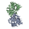
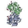
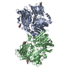
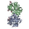
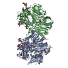
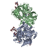
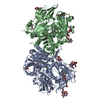
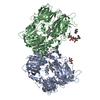
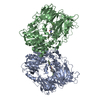
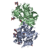
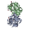
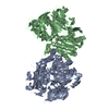
| Entry | Database: PDB / ID: 5y7k | ||||||
|---|---|---|---|---|---|---|---|
| Title | Crystal structure of human DPP4 in complex with inhibitor1 | ||||||
 Components Components | Dipeptidyl peptidase 4 | ||||||
 Keywords Keywords | HYDROLASE / DPP4 / inhibitor | ||||||
| Function / homology |  Function and homology information Function and homology informationglucagon processing / negative regulation of neutrophil chemotaxis / regulation of cell-cell adhesion mediated by integrin / Synthesis, secretion, and inactivation of Glucose-dependent Insulinotropic Polypeptide (GIP) / dipeptidyl-peptidase IV / negative regulation of extracellular matrix disassembly / chemorepellent activity / intercellular canaliculus / psychomotor behavior / dipeptidyl-peptidase activity ...glucagon processing / negative regulation of neutrophil chemotaxis / regulation of cell-cell adhesion mediated by integrin / Synthesis, secretion, and inactivation of Glucose-dependent Insulinotropic Polypeptide (GIP) / dipeptidyl-peptidase IV / negative regulation of extracellular matrix disassembly / chemorepellent activity / intercellular canaliculus / psychomotor behavior / dipeptidyl-peptidase activity / peptide hormone processing / lamellipodium membrane / locomotory exploration behavior / aminopeptidase activity / endocytic vesicle / endothelial cell migration / behavioral fear response / T cell costimulation / receptor-mediated endocytosis of virus by host cell / serine-type peptidase activity / T cell activation / Synthesis, secretion, and inactivation of Glucagon-like Peptide-1 (GLP-1) / lamellipodium / virus receptor activity / protease binding / membrane fusion / response to hypoxia / receptor-mediated virion attachment to host cell / cell adhesion / apical plasma membrane / membrane raft / signaling receptor binding / serine-type endopeptidase activity / lysosomal membrane / focal adhesion / positive regulation of cell population proliferation / symbiont entry into host cell / cell surface / protein homodimerization activity / proteolysis / extracellular exosome / extracellular region / membrane / identical protein binding / plasma membrane Similarity search - Function | ||||||
| Biological species |  Homo sapiens (human) Homo sapiens (human) | ||||||
| Method |  X-RAY DIFFRACTION / SYNCHROTRON / MOLECULAR REPLACEMENT / Resolution: 2.512 Å X-RAY DIFFRACTION / SYNCHROTRON / MOLECULAR REPLACEMENT / Resolution: 2.512 Å | ||||||
 Authors Authors | Lee, H.K. / Kim, E.E. | ||||||
 Citation Citation | Journal: Biochem.Biophys.Res.Commun. / Year: 2017 Title: Unique binding mode of Evogliptin with human dipeptidyl peptidase IV. Authors: Lee, H.K. / Kim, M.K. / Kim, H.D. / Kim, H.J. / Kim, J.W. / Lee, J.O. / Kim, C.W. / Kim, E.E. | ||||||
| History |
|
- Structure visualization
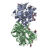
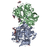
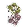
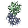
| Structure viewer | Molecule: MolmilJmol/JSmol |
|---|
- Downloads & links
-Download
| PDBx/mmCIF format | 5y7k.cif.gz | 591.8 KB | Display | PDBx/mmCIF format |
|---|---|---|---|---|
| PDB format | pdb5y7k.ent.gz | 488.2 KB | Display | PDB format |
| PDBx/mmJSON format | 5y7k.json.gz | Tree view | PDBx/mmJSON format | |
| Others |  Other downloads Other downloads |
-Validation report
| Arichive directory | https://data.pdbj.org/pub/pdb/validation_reports/y7/5y7kftp://data.pdbj.org/pub/pdb/validation_reports/y7/5y7k | HTTPS FTP |
|---|
-Related structure data
| Related structure data |  5y7hC  5y7jC  1x70S S: Starting model for refinement C: citing same article ( |
|---|---|
| Similar structure data |

-Links
 PDBj
PDBj


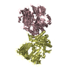
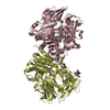
- Assembly
| Deposited unit | 
| ||||||||
|---|---|---|---|---|---|---|---|---|---|
| 1 | 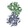
| ||||||||
| 2 | 
| ||||||||
| Unit cell |
|
-Components
| #1: Protein | Mass: 84462.617 Da / Num. of mol.: 4 Source method: isolated from a genetically manipulated source Source: (gene. exp.) Homo sapiens (human) / Gene: DPP4, ADCP2, CD26 / Production host:  Trichoplusia ni (cabbage looper) / References: UniProt: P27487, dipeptidyl-peptidase IV Trichoplusia ni (cabbage looper) / References: UniProt: P27487, dipeptidyl-peptidase IV#2: Chemical | ChemComp-8VU / ( 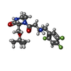  Mass: 401.423 Da / Num. of mol.: 4 / Source method: obtained synthetically / Formula: C19H26F3N3O3 Mass: 401.423 Da / Num. of mol.: 4 / Source method: obtained synthetically / Formula: C19H26F3N3O3#3: Water | ChemComp-HOH / |  Mass: 18.015 Da / Num. of mol.: 272 / Source method: isolated from a natural source / Formula: H2O Mass: 18.015 Da / Num. of mol.: 272 / Source method: isolated from a natural source / Formula: H2OHas protein modification | Y | |
|---|
-Experimental details
-Experiment
| Experiment | Method: X-RAY DIFFRACTION / Number of used crystals: 1 |
|---|
- Sample preparation
| Crystal | Density Matthews: 2.88 Å3/Da / Density % sol: 57.3 % |
|---|---|
| Crystal grow | Temperature: 295 K / Method: vapor diffusion, hanging drop Details: 20~28% (w/v) polyethylene glycol 4000, 0.1M HEPES or Tris pH 7.5~8.5 PH range: 7.5~8.5 |
-Data collection
| Diffraction | Mean temperature: 100 K |
|---|---|
| Diffraction source | Source: SYNCHROTRON / Site: PAL/PLS  / Beamline: 4A / Wavelength: 1 Å / Beamline: 4A / Wavelength: 1 Å |
| Detector | Type: ADSC QUANTUM 210 / Detector: CCD / Date: Oct 18, 2007 |
| Radiation | Protocol: SINGLE WAVELENGTH / Monochromatic (M) / Laue (L): M / Scattering type: x-ray |
| Radiation wavelength | Wavelength: 1 Å / Relative weight: 1 |
| Reflection | Resolution: 2.512→41.136 Å / Num. obs: 129816 / % possible obs: 97.7 % / Redundancy: 2.8 % / Net I/σ(I): 14.1 |
| Reflection shell | Resolution: 2.52→2.61 Å / Redundancy: 1.9 % / Mean I/σ(I) obs: 2.07 / % possible all: 92.3 |
- Processing
| Software |
| |||||||||||||||||||||||||||||||||||||||||||||||||||||||||||||||||||||||||||||||||||||||||||||||||||||||||||||||||||||||||||||||||||||||||||||||||||||||||||||||||||||||||||||||||||||||||||||||||||||||||||||||||||||||||
|---|---|---|---|---|---|---|---|---|---|---|---|---|---|---|---|---|---|---|---|---|---|---|---|---|---|---|---|---|---|---|---|---|---|---|---|---|---|---|---|---|---|---|---|---|---|---|---|---|---|---|---|---|---|---|---|---|---|---|---|---|---|---|---|---|---|---|---|---|---|---|---|---|---|---|---|---|---|---|---|---|---|---|---|---|---|---|---|---|---|---|---|---|---|---|---|---|---|---|---|---|---|---|---|---|---|---|---|---|---|---|---|---|---|---|---|---|---|---|---|---|---|---|---|---|---|---|---|---|---|---|---|---|---|---|---|---|---|---|---|---|---|---|---|---|---|---|---|---|---|---|---|---|---|---|---|---|---|---|---|---|---|---|---|---|---|---|---|---|---|---|---|---|---|---|---|---|---|---|---|---|---|---|---|---|---|---|---|---|---|---|---|---|---|---|---|---|---|---|---|---|---|---|---|---|---|---|---|---|---|---|---|---|---|---|---|---|---|---|
| Refinement | Method to determine structure: MOLECULAR REPLACEMENT Starting model: 1X70 Resolution: 2.512→41.136 Å / SU ML: 0.34 / Cross valid method: FREE R-VALUE / σ(F): 1.44 / Phase error: 24.29 / Stereochemistry target values: ML
| |||||||||||||||||||||||||||||||||||||||||||||||||||||||||||||||||||||||||||||||||||||||||||||||||||||||||||||||||||||||||||||||||||||||||||||||||||||||||||||||||||||||||||||||||||||||||||||||||||||||||||||||||||||||||
| Solvent computation | Shrinkage radii: 0.9 Å / VDW probe radii: 1.11 Å / Solvent model: FLAT BULK SOLVENT MODEL | |||||||||||||||||||||||||||||||||||||||||||||||||||||||||||||||||||||||||||||||||||||||||||||||||||||||||||||||||||||||||||||||||||||||||||||||||||||||||||||||||||||||||||||||||||||||||||||||||||||||||||||||||||||||||
| Refinement step | Cycle: LAST / Resolution: 2.512→41.136 Å
| |||||||||||||||||||||||||||||||||||||||||||||||||||||||||||||||||||||||||||||||||||||||||||||||||||||||||||||||||||||||||||||||||||||||||||||||||||||||||||||||||||||||||||||||||||||||||||||||||||||||||||||||||||||||||
| Refine LS restraints |
| |||||||||||||||||||||||||||||||||||||||||||||||||||||||||||||||||||||||||||||||||||||||||||||||||||||||||||||||||||||||||||||||||||||||||||||||||||||||||||||||||||||||||||||||||||||||||||||||||||||||||||||||||||||||||
| LS refinement shell |
|
