 Movie
Movie Controller

[English] 日本語
 Yorodumi
- PDB-5y3z: Structure of the periplasmic domain of the MotB L119P mutant from... -
+ Open data

- Basic information
| Entry | Database: PDB / ID: 5y3z | ||||||
|---|---|---|---|---|---|---|---|
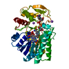
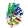
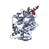
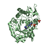
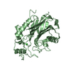
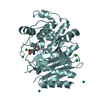
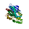
| Title | Structure of the periplasmic domain of the MotB L119P mutant from Salmonella (crystal form 1) | ||||||
 Components Components | Motility protein B | ||||||
 Keywords Keywords | MOTOR PROTEIN / 2-LAYER SANDWICH / BACTERIAL FLAGELLUM / CELL PROJECTION / CHEMOTAXIS / FLAGELLAR ROTATION / INNER MEMBRANE / MEMBRANE PROTEIN | ||||||
| Function / homology |  Function and homology information Function and homology informationbacterial-type flagellum stator complex / bacterial-type flagellum-dependent cell motility / chemotaxis Similarity search - Function | ||||||
| Biological species |  Salmonella typhimurium (bacteria) Salmonella typhimurium (bacteria) | ||||||
| Method |  X-RAY DIFFRACTION / SYNCHROTRON / MOLECULAR REPLACEMENT / Resolution: 2 Å X-RAY DIFFRACTION / SYNCHROTRON / MOLECULAR REPLACEMENT / Resolution: 2 Å | ||||||
 Authors Authors | Takao, M. / Kojima, S. / Sakuma, M. / Homma, M. / Imada, K. | ||||||
| Funding support |  Japan, 1items Japan, 1items
| ||||||
 Citation Citation | Journal: Structure / Year: 2018 Title: The Helix Rearrangement in the Periplasmic Domain of the Flagellar Stator B Subunit Activates Peptidoglycan Binding and Ion Influx. Authors: Kojima, S. / Takao, M. / Almira, G. / Kawahara, I. / Sakuma, M. / Homma, M. / Kojima, C. / Imada, K. | ||||||
| History |
|
- Structure visualization
| Structure viewer | Molecule: MolmilJmol/JSmol |
|---|
- Downloads & links
-Download
| PDBx/mmCIF format | 5y3z.cif.gz | 76.7 KB | Display | PDBx/mmCIF format |
|---|---|---|---|---|
| PDB format | pdb5y3z.ent.gz | 55.1 KB | Display | PDB format |
| PDBx/mmJSON format | 5y3z.json.gz | Tree view | PDBx/mmJSON format | |
| Others |  Other downloads Other downloads |
-Validation report
| Arichive directory | https://data.pdbj.org/pub/pdb/validation_reports/y3/5y3zftp://data.pdbj.org/pub/pdb/validation_reports/y3/5y3z | HTTPS FTP |
|---|
-Related structure data
| Related structure data |  5y40C  2zvyS S: Starting model for refinement C: citing same article ( |
|---|---|
| Similar structure data |


-Links
 PDBj
PDBj


- Assembly
| Deposited unit | 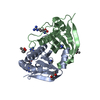
| ||||||||
|---|---|---|---|---|---|---|---|---|---|
| 1 |
| ||||||||
| Unit cell |
|
-Components
| #1: Protein | Mass: 20718.629 Da / Num. of mol.: 2 / Fragment: UNP residues 99-276 / Mutation: L119P Source method: isolated from a genetically manipulated source Source: (gene. exp.) Salmonella typhimurium (strain LT2 / SGSC1412 / ATCC 700720) (bacteria)Strain: LT2 / SGSC1412 / ATCC 700720 / Gene: motB, STM1922 / Variant: SJW1103 / Plasmid: PET19b / Production host: #2: Chemical |   Type: L-peptide linking / Mass: 175.209 Da / Num. of mol.: 3 / Source method: obtained synthetically / Formula: C6H15N4O2 Type: L-peptide linking / Mass: 175.209 Da / Num. of mol.: 3 / Source method: obtained synthetically / Formula: C6H15N4O2#3: Chemical |   Mass: 69.085 Da / Num. of mol.: 2 / Source method: obtained synthetically / Formula: C3H5N2 Mass: 69.085 Da / Num. of mol.: 2 / Source method: obtained synthetically / Formula: C3H5N2#4: Chemical |   Mass: 118.174 Da / Num. of mol.: 3 / Source method: obtained synthetically / Formula: C6H14O2 / Comment: precipitant*YM Mass: 118.174 Da / Num. of mol.: 3 / Source method: obtained synthetically / Formula: C6H14O2 / Comment: precipitant*YM#5: Water | ChemComp-HOH / |  Mass: 18.015 Da / Num. of mol.: 151 / Source method: isolated from a natural source / Formula: H2O Mass: 18.015 Da / Num. of mol.: 151 / Source method: isolated from a natural source / Formula: H2O |
|---|
-Experimental details
-Experiment
| Experiment | Method: X-RAY DIFFRACTION / Number of used crystals: 1 |
|---|
- Sample preparation
| Crystal | Density Matthews: 1.96 Å3/Da / Density % sol: 37.21 % |
|---|---|
| Crystal grow | Temperature: 293 K / Method: vapor diffusion, sitting drop / pH: 6.5 Details: 28% PEG 3350, 0.1M imidazole-HCl, 0.15M Arg-HCl, 5% MPD |
-Data collection
| Diffraction | Mean temperature: 100 K |
|---|---|
| Diffraction source | Source: SYNCHROTRON / Site: SPring-8 / Beamline: BL41XU / Wavelength: 1 Å |
| Detector | Type: MARMOSAIC 225 mm CCD / Detector: CCD / Date: Nov 1, 2010 |
| Radiation | Monochromator: Double-crystal monochromator / Protocol: SINGLE WAVELENGTH / Monochromatic (M) / Laue (L): M / Scattering type: x-ray |
| Radiation wavelength | Wavelength: 1 Å / Relative weight: 1 |
| Reflection | Resolution: 2→43.4 Å / Num. obs: 22684 / % possible obs: 99.9 % / Redundancy: 4.1 % / Biso Wilson estimate: 18.7 Å2 / Rmerge(I) obs: 0.093 / Net I/σ(I): 10.7 |
| Reflection shell | Resolution: 2→2.05 Å / Redundancy: 4.1 % / Rmerge(I) obs: 0.496 / Mean I/σ(I) obs: 2.7 / Num. unique obs: 1629 / % possible all: 100 |
- Processing
| Software |
| |||||||||||||||||||||||||||||||||||||||||||||||||||||||||||||||||||||||||||||||||||||||||||||||||||||||||
|---|---|---|---|---|---|---|---|---|---|---|---|---|---|---|---|---|---|---|---|---|---|---|---|---|---|---|---|---|---|---|---|---|---|---|---|---|---|---|---|---|---|---|---|---|---|---|---|---|---|---|---|---|---|---|---|---|---|---|---|---|---|---|---|---|---|---|---|---|---|---|---|---|---|---|---|---|---|---|---|---|---|---|---|---|---|---|---|---|---|---|---|---|---|---|---|---|---|---|---|---|---|---|---|---|---|---|
| Refinement | Method to determine structure: MOLECULAR REPLACEMENT Starting model: 2ZVY Resolution: 2→35.687 Å / SU ML: 0.2 / Cross valid method: FREE R-VALUE / σ(F): 1.35 / Phase error: 20.38 / Stereochemistry target values: ML
| |||||||||||||||||||||||||||||||||||||||||||||||||||||||||||||||||||||||||||||||||||||||||||||||||||||||||
| Solvent computation | Shrinkage radii: 0.9 Å / VDW probe radii: 1.11 Å / Solvent model: FLAT BULK SOLVENT MODEL | |||||||||||||||||||||||||||||||||||||||||||||||||||||||||||||||||||||||||||||||||||||||||||||||||||||||||
| Refinement step | Cycle: LAST / Resolution: 2→35.687 Å
| |||||||||||||||||||||||||||||||||||||||||||||||||||||||||||||||||||||||||||||||||||||||||||||||||||||||||
| Refine LS restraints |
| |||||||||||||||||||||||||||||||||||||||||||||||||||||||||||||||||||||||||||||||||||||||||||||||||||||||||
| LS refinement shell |
|
