 Movie
Movie Controller

[English] 日本語
 Yorodumi
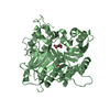
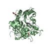
- PDB-5xyh: Crystal Structure of catalytic domain of 1,4-beta-Cellobiosidase ... -
+ Open data

- Basic information
| Entry | Database: PDB / ID: 5xyh | ||||||
|---|---|---|---|---|---|---|---|
| Title | Crystal Structure of catalytic domain of 1,4-beta-Cellobiosidase (CbsA) from Xanthomonas oryzae pv. oryzae | ||||||
 Components Components | CbsA | ||||||
 Keywords Keywords | HYDROLASE / exoglucanase / Xoo / cell-wall degrading enzyme / bacterial blight | ||||||
| Function / homology |  Function and homology information Function and homology informationHydrolases; Glycosylases; Glycosidases, i.e. enzymes that hydrolyse O- and S-glycosyl compounds / hydrolase activity, hydrolyzing O-glycosyl compounds / cellulose catabolic process Similarity search - Function | ||||||
| Biological species |  Xanthomonas oryzae pv. oryzae (bacteria) Xanthomonas oryzae pv. oryzae (bacteria) | ||||||
| Method |  X-RAY DIFFRACTION / MOLECULAR REPLACEMENT / Resolution: 1.864 Å X-RAY DIFFRACTION / MOLECULAR REPLACEMENT / Resolution: 1.864 Å | ||||||
 Authors Authors | Kumar, S. / Haque, A.S. / Nathawat, R. / Sankaranaryanan, R. | ||||||
| Funding support |  India, 1items India, 1items
| ||||||
 Citation Citation | Journal: Mol. Plant Pathol. / Year: 2018 Title: A mutation in an exoglucanase of Xanthomonas oryzae pv. oryzae, which confers an endo mode of activity, affects bacterial virulence, but not the induction of immune responses, in rice Authors: Tayi, L. / Kumar, S. / Nathawat, R. / Haque, A.S. / Maku, R.V. / Patel, H.K. / Sankaranarayanan, R. / Sonti, R.V. #1: Journal: Acta Crystallogr. Sect. F Struct. Biol. Cryst. Commun. Year: 2012 Title: Crystallization and preliminary crystallographic studies of CbsA, a secretory exoglucanase from Xanthomonas oryzae pv. oryzae. Authors: Kumar, S. / Haque, A.S. / Jha, G. / Sonti, R.V. / Sankaranarayanan, R. | ||||||
| History |
|
- Structure visualization
| Structure viewer | Molecule: MolmilJmol/JSmol |
|---|
- Downloads & links
-Download
| PDBx/mmCIF format | 5xyh.cif.gz | 103.5 KB | Display | PDBx/mmCIF format |
|---|---|---|---|---|
| PDB format | pdb5xyh.ent.gz | 76.1 KB | Display | PDB format |
| PDBx/mmJSON format | 5xyh.json.gz | Tree view | PDBx/mmJSON format | |
| Others |  Other downloads Other downloads |
-Validation report
| Arichive directory | https://data.pdbj.org/pub/pdb/validation_reports/xy/5xyhftp://data.pdbj.org/pub/pdb/validation_reports/xy/5xyh | HTTPS FTP |
|---|
-Related structure data
| Related structure data |  4b4hS S: Starting model for refinement |
|---|---|
| Similar structure data |


-Links
 PDBj
PDBj- Assembly
| Deposited unit | 
| ||||||||
|---|---|---|---|---|---|---|---|---|---|
| 1 |
| ||||||||
| Unit cell |
|
-Components
| #1: Protein | Mass: 45624.789 Da / Num. of mol.: 1 Source method: isolated from a genetically manipulated source Details: culture supernatant Source: (gene. exp.) Xanthomonas oryzae pv. oryzae (bacteria)Strain: BXO43 / Gene: cbsA / Plasmid: pUFR034 / Details (production host): overexpression / Production host: Xanthomonas oryzae pv. oryzae (bacteria) / Strain (production host): BXO2700References: UniProt: A0A384E106*PLUS, cellulose 1,4-beta-cellobiosidase (non-reducing end) |
|---|---|
| #2: Water | ChemComp-HOH /  Mass: 18.015 Da / Num. of mol.: 442 / Source method: isolated from a natural source / Formula: H2O Mass: 18.015 Da / Num. of mol.: 442 / Source method: isolated from a natural source / Formula: H2O |
| Has protein modification | Y |
-Experimental details
-Experiment
| Experiment | Method: X-RAY DIFFRACTION / Number of used crystals: 1 |
|---|
- Sample preparation
| Crystal | Density Matthews: 2.29 Å3/Da / Density % sol: 46.26 % |
|---|---|
| Crystal grow | Temperature: 277 K / Method: vapor diffusion, hanging drop / Details: polyethylene glycol 3350 citric acid / PH range: 3.0 to 6.5 |
-Data collection
| Diffraction | Mean temperature: 100 K |
|---|---|
| Diffraction source | Source: ROTATING ANODE / Type: RIGAKU MICROMAX-007 HF / Wavelength: 1.54179 Å |
| Detector | Type: MAR scanner 345 mm plate / Detector: IMAGE PLATE / Date: Nov 28, 2011 |
| Radiation | Protocol: SINGLE WAVELENGTH / Monochromatic (M) / Laue (L): M / Scattering type: x-ray |
| Radiation wavelength | Wavelength: 1.54179 Å / Relative weight: 1 |
| Reflection | Resolution: 1.86→25 Å / Num. obs: 33848 / % possible obs: 93.9 % / Redundancy: 6.7 % / Rmerge(I) obs: 0.049 / Net I/σ(I): 31.69 |
| Reflection shell | Resolution: 1.86→1.93 Å / Redundancy: 5.6 % / Rmerge(I) obs: 0.183 / Mean I/σ(I) obs: 7.75 / % possible all: 89.4 |
- Processing
| Software |
| |||||||||||||||||||||||||||||||||||||||||||||||||||||||||||||||||||||||||||||||||||||||||||
|---|---|---|---|---|---|---|---|---|---|---|---|---|---|---|---|---|---|---|---|---|---|---|---|---|---|---|---|---|---|---|---|---|---|---|---|---|---|---|---|---|---|---|---|---|---|---|---|---|---|---|---|---|---|---|---|---|---|---|---|---|---|---|---|---|---|---|---|---|---|---|---|---|---|---|---|---|---|---|---|---|---|---|---|---|---|---|---|---|---|---|---|---|
| Refinement | Method to determine structure: MOLECULAR REPLACEMENT Starting model: 4B4H Resolution: 1.864→24.517 Å / SU ML: 0.23 / Cross valid method: FREE R-VALUE / σ(F): 1.33 / Phase error: 24.04 / Stereochemistry target values: ML
| |||||||||||||||||||||||||||||||||||||||||||||||||||||||||||||||||||||||||||||||||||||||||||
| Solvent computation | Shrinkage radii: 0.9 Å / VDW probe radii: 1.11 Å / Solvent model: FLAT BULK SOLVENT MODEL | |||||||||||||||||||||||||||||||||||||||||||||||||||||||||||||||||||||||||||||||||||||||||||
| Refinement step | Cycle: LAST / Resolution: 1.864→24.517 Å
| |||||||||||||||||||||||||||||||||||||||||||||||||||||||||||||||||||||||||||||||||||||||||||
| Refine LS restraints |
| |||||||||||||||||||||||||||||||||||||||||||||||||||||||||||||||||||||||||||||||||||||||||||
| LS refinement shell |
|
