 Movie
Movie Controller

[English] 日本語
 Yorodumi
- PDB-5xtr: Crystal structure of baculoviral sulfhydryl oxidase P33 (R127A, E... -
+ Open data

- Basic information
| Entry | Database: PDB / ID: 5xtr | ||||||
|---|---|---|---|---|---|---|---|
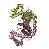
| Title | Crystal structure of baculoviral sulfhydryl oxidase P33 (R127A, E183A mutant) | ||||||
 Components Components | FAD-linked sulfhydryl oxidase | ||||||
 Keywords Keywords | OXIDOREDUCTASE / sulfhydryl oxidase | ||||||
| Function / homology | FAD-linked sulfhydryl oxidase / Baculovirus P33 / thiol oxidase activity / thiol oxidase / host cell cytoplasm / host cell nucleus / FLAVIN-ADENINE DINUCLEOTIDE / FAD-linked sulfhydryl oxidase Function and homology information Function and homology information | ||||||
| Biological species |  Autographa californica nuclear polyhedrosis virus Autographa californica nuclear polyhedrosis virus | ||||||
| Method |  X-RAY DIFFRACTION / SYNCHROTRON / MOLECULAR REPLACEMENT / Resolution: 2.25 Å X-RAY DIFFRACTION / SYNCHROTRON / MOLECULAR REPLACEMENT / Resolution: 2.25 Å | ||||||
 Authors Authors | Kuang, W. / Hu, Z. / Gong, P. | ||||||
| Funding support |  China, 1items China, 1items
| ||||||
 Citation Citation | Journal: J. Virol. / Year: 2017 Title: Three Conserved Regions in Baculovirus Sulfhydryl Oxidase P33 Are Critical for Enzymatic Activity and Function Authors: Kuang, W. / Zhang, H. / Wang, M. / Zhou, N.Y. / Deng, F. / Wang, H. / Gong, P. / Hu, Z. | ||||||
| History |
|
- Structure visualization
| Structure viewer | Molecule: MolmilJmol/JSmol |
|---|
- Downloads & links
-Download
| PDBx/mmCIF format | 5xtr.cif.gz | 216.9 KB | Display | PDBx/mmCIF format |
|---|---|---|---|---|
| PDB format | pdb5xtr.ent.gz | 170.7 KB | Display | PDB format |
| PDBx/mmJSON format | 5xtr.json.gz | Tree view | PDBx/mmJSON format | |
| Others |  Other downloads Other downloads |
-Validation report
| Arichive directory | https://data.pdbj.org/pub/pdb/validation_reports/xt/5xtrftp://data.pdbj.org/pub/pdb/validation_reports/xt/5xtr | HTTPS FTP |
|---|
-Related structure data
| Related structure data | 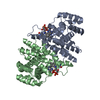 5xkiC 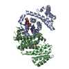 5xtnC 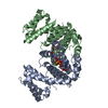 5xtoC  5xtpC 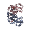 5xtqC  3qzyS S: Starting model for refinement C: citing same article ( |
|---|---|
| Similar structure data |


-Links
 PDBj
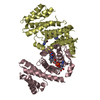
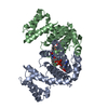
PDBj- Assembly
| Deposited unit | 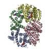
| ||||||||
|---|---|---|---|---|---|---|---|---|---|
| 1 | 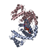
| ||||||||
| 2 | 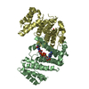
| ||||||||
| Unit cell |
|
-Components
| #1: Protein | Mass: 34382.789 Da / Num. of mol.: 4 / Mutation: R127A, E183A Source method: isolated from a genetically manipulated source Source: (gene. exp.) Autographa californica nuclear polyhedrosis virusGene: P33, ORF92 / Plasmid: pET28 / Production host:  #2: Chemical | ChemComp-FAD /   Mass: 785.550 Da / Num. of mol.: 4 / Source method: obtained synthetically / Formula: C27H33N9O15P2 / Comment: FAD*YM Mass: 785.550 Da / Num. of mol.: 4 / Source method: obtained synthetically / Formula: C27H33N9O15P2 / Comment: FAD*YM#3: Water | ChemComp-HOH / |  Mass: 18.015 Da / Num. of mol.: 246 / Source method: isolated from a natural source / Formula: H2O Mass: 18.015 Da / Num. of mol.: 246 / Source method: isolated from a natural source / Formula: H2OHas protein modification | Y | |
|---|
-Experimental details
-Experiment
| Experiment | Method: X-RAY DIFFRACTION / Number of used crystals: 1 |
|---|
- Sample preparation
| Crystal | Density Matthews: 2.32 Å3/Da / Density % sol: 47.03 % |
|---|---|
| Crystal grow | Temperature: 293 K / Method: vapor diffusion, sitting drop / pH: 4.3 / Details: PEG3350 |
-Data collection
| Diffraction | Mean temperature: 100 K | ||||||||||||||||||||||||||||||||||||||||||||||||||||||||||||||||||||||||||||||||||||||||
|---|---|---|---|---|---|---|---|---|---|---|---|---|---|---|---|---|---|---|---|---|---|---|---|---|---|---|---|---|---|---|---|---|---|---|---|---|---|---|---|---|---|---|---|---|---|---|---|---|---|---|---|---|---|---|---|---|---|---|---|---|---|---|---|---|---|---|---|---|---|---|---|---|---|---|---|---|---|---|---|---|---|---|---|---|---|---|---|---|---|
| Diffraction source | Source: SYNCHROTRON / Site: SSRF / Beamline: BL17U1 / Wavelength: 0.9789 Å | ||||||||||||||||||||||||||||||||||||||||||||||||||||||||||||||||||||||||||||||||||||||||
| Detector | Type: ADSC QUANTUM 315r / Detector: CCD / Date: Nov 18, 2014 | ||||||||||||||||||||||||||||||||||||||||||||||||||||||||||||||||||||||||||||||||||||||||
| Radiation | Protocol: SINGLE WAVELENGTH / Monochromatic (M) / Laue (L): M / Scattering type: x-ray | ||||||||||||||||||||||||||||||||||||||||||||||||||||||||||||||||||||||||||||||||||||||||
| Radiation wavelength | Wavelength: 0.9789 Å / Relative weight: 1 | ||||||||||||||||||||||||||||||||||||||||||||||||||||||||||||||||||||||||||||||||||||||||
| Reflection | Resolution: 2.25→37.91 Å / Num. obs: 53132 / % possible obs: 90.5 % / Redundancy: 1.83 % / Biso Wilson estimate: 37.97 Å2 / Rmerge(I) obs: 0.057 / Rrim(I) all: 0.081 / Χ2: 1.22 / Net I/σ(I): 7.4 | ||||||||||||||||||||||||||||||||||||||||||||||||||||||||||||||||||||||||||||||||||||||||
| Reflection shell | Diffraction-ID: 1
|
- Processing
| Software |
| ||||||||||||||||||||||||||||||||||||||||||||||||||||||||||||||||||||||||||||||||||||||||||||||||||||||||||||||||||||||||||||||
|---|---|---|---|---|---|---|---|---|---|---|---|---|---|---|---|---|---|---|---|---|---|---|---|---|---|---|---|---|---|---|---|---|---|---|---|---|---|---|---|---|---|---|---|---|---|---|---|---|---|---|---|---|---|---|---|---|---|---|---|---|---|---|---|---|---|---|---|---|---|---|---|---|---|---|---|---|---|---|---|---|---|---|---|---|---|---|---|---|---|---|---|---|---|---|---|---|---|---|---|---|---|---|---|---|---|---|---|---|---|---|---|---|---|---|---|---|---|---|---|---|---|---|---|---|---|---|---|
| Refinement | Method to determine structure: MOLECULAR REPLACEMENT Starting model: 3QZY Resolution: 2.25→37.883 Å / SU ML: 0.32 / Cross valid method: FREE R-VALUE / σ(F): 1.96 / Phase error: 33.33
| ||||||||||||||||||||||||||||||||||||||||||||||||||||||||||||||||||||||||||||||||||||||||||||||||||||||||||||||||||||||||||||||
| Solvent computation | Shrinkage radii: 0.9 Å / VDW probe radii: 1.11 Å | ||||||||||||||||||||||||||||||||||||||||||||||||||||||||||||||||||||||||||||||||||||||||||||||||||||||||||||||||||||||||||||||
| Displacement parameters | Biso max: 87.96 Å2 / Biso mean: 41.3415 Å2 / Biso min: 20.49 Å2 | ||||||||||||||||||||||||||||||||||||||||||||||||||||||||||||||||||||||||||||||||||||||||||||||||||||||||||||||||||||||||||||||
| Refinement step | Cycle: final / Resolution: 2.25→37.883 Å
| ||||||||||||||||||||||||||||||||||||||||||||||||||||||||||||||||||||||||||||||||||||||||||||||||||||||||||||||||||||||||||||||
| Refine LS restraints |
| ||||||||||||||||||||||||||||||||||||||||||||||||||||||||||||||||||||||||||||||||||||||||||||||||||||||||||||||||||||||||||||||
| LS refinement shell | Refine-ID: X-RAY DIFFRACTION / Rfactor Rfree error: 0 / Total num. of bins used: 17
|
