 Movie
Movie Controller

[English] 日本語
 Yorodumi
- PDB-5xpt: Crystal structure of MAD2L2/REV7 in complex with a CAMP fragment ... -
+ Open data

- Basic information
| Entry | Database: PDB / ID: 5xpt | ||||||
|---|---|---|---|---|---|---|---|
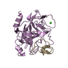
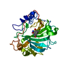
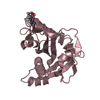
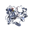
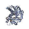
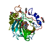
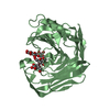
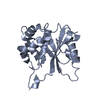
| Title | Crystal structure of MAD2L2/REV7 in complex with a CAMP fragment in a tetragonal crystal | ||||||
 Components Components |
| ||||||
 Keywords Keywords | TRANSCRIPTION/METAL BINDING PROTEIN / MAD2L2 / MAD2B / REV7 / CAMP / champ1 / TRANSCRIPTION-METAL BINDING PROTEIN complex | ||||||
| Function / homology |  Function and homology information Function and homology informationsomatic diversification of immunoglobulins involved in immune response / DNA damage response, signal transduction resulting in transcription / negative regulation of ubiquitin protein ligase activity / zeta DNA polymerase complex / sister chromatid biorientation / protein localization to microtubule / positive regulation of isotype switching / : / JUN kinase binding / attachment of mitotic spindle microtubules to kinetochore ...somatic diversification of immunoglobulins involved in immune response / DNA damage response, signal transduction resulting in transcription / negative regulation of ubiquitin protein ligase activity / zeta DNA polymerase complex / sister chromatid biorientation / protein localization to microtubule / positive regulation of isotype switching / : / JUN kinase binding / attachment of mitotic spindle microtubules to kinetochore / negative regulation of cell-cell adhesion mediated by cadherin / protein localization to kinetochore / negative regulation of epithelial to mesenchymal transition / positive regulation of double-strand break repair via nonhomologous end joining / mitotic spindle assembly checkpoint signaling / telomere maintenance in response to DNA damage / positive regulation of peptidyl-serine phosphorylation / error-prone translesion synthesis / negative regulation of double-strand break repair via homologous recombination / site of DNA damage / condensed chromosome / translesion synthesis / Translesion synthesis by REV1 / Translesion synthesis by POLK / Translesion synthesis by POLI / actin filament organization / regulation of cell growth / negative regulation of canonical Wnt signaling pathway / negative regulation of protein catabolic process / kinetochore / spindle / transcription corepressor activity / double-strand break repair / chromosome / site of double-strand break / RNA polymerase II-specific DNA-binding transcription factor binding / protein-macromolecule adaptor activity / nuclear body / cell division / DNA repair / positive regulation of DNA-templated transcription / chromatin / negative regulation of transcription by RNA polymerase II / zinc ion binding / nucleoplasm / nucleus / cytoplasm Similarity search - Function | ||||||
| Biological species |  Homo sapiens (human) Homo sapiens (human) | ||||||
| Method |  X-RAY DIFFRACTION / SYNCHROTRON / MOLECULAR REPLACEMENT / Resolution: 2.102 Å X-RAY DIFFRACTION / SYNCHROTRON / MOLECULAR REPLACEMENT / Resolution: 2.102 Å | ||||||
 Authors Authors | Hara, K. / Taharazako, S. / Hashimoto, H. | ||||||
 Citation Citation | Journal: J. Biol. Chem. / Year: 2017 Title: Dynamic feature of mitotic arrest deficient 2-like protein 2 (MAD2L2) and structural basis for its interaction with chromosome alignment-maintaining phosphoprotein (CAMP). Authors: Hara, K. / Taharazako, S. / Ikeda, M. / Fujita, H. / Mikami, Y. / Kikuchi, S. / Hishiki, A. / Yokoyama, H. / Ishikawa, Y. / Kanno, S.I. / Tanaka, K. / Hashimoto, H. | ||||||
| History |
|
- Structure visualization
| Structure viewer | Molecule: MolmilJmol/JSmol |
|---|
- Downloads & links
-Download
| PDBx/mmCIF format | 5xpt.cif.gz | 60.5 KB | Display | PDBx/mmCIF format |
|---|---|---|---|---|
| PDB format | pdb5xpt.ent.gz | 42.1 KB | Display | PDB format |
| PDBx/mmJSON format | 5xpt.json.gz | Tree view | PDBx/mmJSON format | |
| Others |  Other downloads Other downloads |
-Validation report
| Arichive directory | https://data.pdbj.org/pub/pdb/validation_reports/xp/5xptftp://data.pdbj.org/pub/pdb/validation_reports/xp/5xpt | HTTPS FTP |
|---|
-Related structure data
| Related structure data | 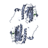 5xpuC  3abeS S: Starting model for refinement C: citing same article ( |
|---|---|
| Similar structure data |


-Links
 PDBj
PDBj


- Assembly
| Deposited unit | 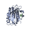
| ||||||||
|---|---|---|---|---|---|---|---|---|---|
| 1 |
| ||||||||
| Unit cell |
| ||||||||
| Components on special symmetry positions |
|
-Components
| #1: Protein | Mass: 26101.236 Da / Num. of mol.: 1 / Mutation: R124A Source method: isolated from a genetically manipulated source Source: (gene. exp.) Homo sapiens (human) / Gene: MAD2L2, MAD2B, REV7 / Production host:  |
|---|---|
| #2: Protein/peptide | Mass: 2085.340 Da / Num. of mol.: 1 / Fragment: UNP RESIDUES 325-344 Source method: isolated from a genetically manipulated source Source: (gene. exp.) Homo sapiens (human) / Gene: CHAMP1, C13orf8, CAMP, CHAMP, KIAA1802, ZNF828 / Production host: |
| #3: Water | ChemComp-HOH /  Mass: 18.015 Da / Num. of mol.: 120 / Source method: isolated from a natural source / Formula: H2O Mass: 18.015 Da / Num. of mol.: 120 / Source method: isolated from a natural source / Formula: H2O |
-Experimental details
-Experiment
| Experiment | Method: X-RAY DIFFRACTION / Number of used crystals: 1 |
|---|
- Sample preparation
| Crystal | Density Matthews: 3.2 Å3/Da / Density % sol: 61.62 % |
|---|---|
| Crystal grow | Temperature: 293 K / Method: vapor diffusion, hanging drop / pH: 6.6 Details: 0.1 M sodium cacodylate pH 6.6, 4.0 M sodium formate |
-Data collection
| Diffraction | Mean temperature: 100 K |
|---|---|
| Diffraction source | Source: SYNCHROTRON / Site: Photon Factory  / Beamline: BL-17A / Wavelength: 0.98 Å / Beamline: BL-17A / Wavelength: 0.98 Å |
| Detector | Type: DECTRIS PILATUS3 S 6M / Detector: PIXEL / Date: Jun 22, 2016 |
| Radiation | Protocol: SINGLE WAVELENGTH / Monochromatic (M) / Laue (L): M / Scattering type: x-ray |
| Radiation wavelength | Wavelength: 0.98 Å / Relative weight: 1 |
| Reflection | Resolution: 2.1→19.85 Å / Num. obs: 21579 / % possible obs: 99.8 % / Redundancy: 13.1 % / Net I/σ(I): 27.5 |
| Reflection shell | Resolution: 2.1→2.22 Å |
- Processing
| Software |
| |||||||||||||||||||||||||||||||||||||||||||||||||||||||||||||||
|---|---|---|---|---|---|---|---|---|---|---|---|---|---|---|---|---|---|---|---|---|---|---|---|---|---|---|---|---|---|---|---|---|---|---|---|---|---|---|---|---|---|---|---|---|---|---|---|---|---|---|---|---|---|---|---|---|---|---|---|---|---|---|---|---|
| Refinement | Method to determine structure: MOLECULAR REPLACEMENT Starting model: 3ABE Resolution: 2.102→19.846 Å / SU ML: 0.21 / Cross valid method: FREE R-VALUE / σ(F): 1.34 / Phase error: 23.23
| |||||||||||||||||||||||||||||||||||||||||||||||||||||||||||||||
| Solvent computation | Shrinkage radii: 0.9 Å / VDW probe radii: 1.11 Å | |||||||||||||||||||||||||||||||||||||||||||||||||||||||||||||||
| Refinement step | Cycle: LAST / Resolution: 2.102→19.846 Å
| |||||||||||||||||||||||||||||||||||||||||||||||||||||||||||||||
| Refine LS restraints |
| |||||||||||||||||||||||||||||||||||||||||||||||||||||||||||||||
| LS refinement shell |
|
