Type: MARMOSAIC 325 mm CCD / Detector: CCD / Date: Nov 11, 2010
Radiation
Protocol: SINGLE WAVELENGTH / Monochromatic (M) / Laue (L): M / Scattering type: x-ray
Radiation wavelength
Wavelength: 0.979 Å / Relative weight: 1
Reflection
Resolution: 2.47→50 Å / Num. obs: 18539 / % possible obs: 99.1 % / Redundancy: 11.6 % / Net I/σ(I): 21.3
-
Processing
Software
Name
Version
Classification
REFMAC
5.7.0032
refinement
HKL-2000
datareduction
HKL-2000
datascaling
PHASER
phasing
Refinement
Resolution: 2.48→49.29 Å / Cor.coef. Fo:Fc: 0.941 / Cor.coef. Fo:Fc free: 0.933 / SU B: 14.395 / SU ML: 0.274 / Cross valid method: THROUGHOUT / ESU R: 0.483 / ESU R Free: 0.311 / Stereochemistry target values: MAXIMUM LIKELIHOOD / Details: HYDROGENS HAVE BEEN ADDED IN THE RIDING POSITIONS
Rfactor
Num. reflection
% reflection
Selection details
Rfree
0.29439
962
5.22 %
RANDOM
Rwork
0.24465
-
-
-
obs
0.24518
18414
98.02 %
-
Solvent computation
Ion probe radii: 0.8 Å / Shrinkage radii: 0.8 Å / VDW probe radii: 1.2 Å / Solvent model: MASK
 Movie
Movie Controller


 Open data
 Basic information
 Components
 Keywords
 Function and homology information
 Archaeoglobus fulgidus (archaea)
 X-RAY DIFFRACTION /
  Authors
 China, 1items
  Citation
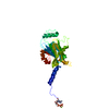
 Structure visualization
 Downloads & links
 Other downloads
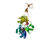


 PDBj
PDBj

 Assembly


 Mass: 65.409 Da / Num. of mol.: 1 / Source method: obtained synthetically / Formula: Zn
 Mass: 24.305 Da / Num. of mol.: 1 / Source method: obtained synthetically / Formula: Mg
 Sample preparation
 Processing