[English] 日本語
Yorodumi
- PDB-5xnh: Crystal structure of the branched-chain polyamine synthase (BpsA)... -

+
Open data


ID or keywords:

Loading...

-
Basic information

Entry
Database: PDB / ID: 5xnh
TitleCrystal structure of the branched-chain polyamine synthase (BpsA) in complex with spermidine
ComponentsN(4)-bis(aminopropyl)spermidine synthase
KeywordsTRANSFERASE / Polyamine biosynthesis / N(4)-bis(aminopropyl)spermidine synthase
Function / homology
Function and homology information


N4-bis(aminopropyl)spermidine synthase / rRNA (adenine-N6-)-methyltransferase activity / polyamine biosynthetic process / transferase activity, transferring alkyl or aryl (other than methyl) groups / transferase activity / cytoplasm
Similarity search - Function
N(4)-bis(aminopropyl)spermidine synthase, C-terminal / N(4)-bis(aminopropyl)spermidine synthase / Branched-chain polyamine synthase A C-terminal domain / Vaccinia Virus protein VP39 / Winged helix-like DNA-binding domain superfamily/Winged helix DNA-binding domain / Arc Repressor Mutant, subunit A / Winged helix-like DNA-binding domain superfamily / S-adenosyl-L-methionine-dependent methyltransferase superfamily / Rossmann fold / Orthogonal Bundle ...N(4)-bis(aminopropyl)spermidine synthase, C-terminal / N(4)-bis(aminopropyl)spermidine synthase / Branched-chain polyamine synthase A C-terminal domain / Vaccinia Virus protein VP39 / Winged helix-like DNA-binding domain superfamily/Winged helix DNA-binding domain / Arc Repressor Mutant, subunit A / Winged helix-like DNA-binding domain superfamily / S-adenosyl-L-methionine-dependent methyltransferase superfamily / Rossmann fold / Orthogonal Bundle / 3-Layer(aba) Sandwich / Mainly Alpha / Alpha Beta
Similarity search - Domain/homology
: / SPERMIDINE / N(4)-bis(aminopropyl)spermidine synthase
Similarity search - Component
Biological species
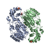
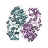
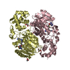
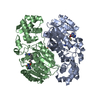
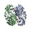
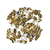
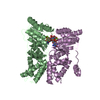
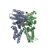
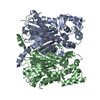
Thermococcus kodakarensis (archaea)
MethodX-RAY DIFFRACTION / SYNCHROTRON / Resolution: 1.95 Å
AuthorsMizohata, E. / Tse, K.M. / Fujita, J. / Inoue, T.
CitationJournal: FEBS J. / Year: 2017
Title: Active site geometry of a novel aminopropyltransferase for biosynthesis of hyperthermophile-specific branched-chain polyamine.
Authors: Hidese, R. / Tse, K.M. / Kimura, S. / Mizohata, E. / Fujita, J. / Horai, Y. / Umezawa, N. / Higuchi, T. / Niitsu, M. / Oshima, T. / Imanaka, T. / Inoue, T. / Fujiwara, S.
History
DepositionMay 22, 2017Deposition site: PDBJ / Processing site: PDBJ
Revision 1.0Oct 4, 2017Provider: repository / Type: Initial release
Revision 1.1Dec 6, 2017Group: Database references / Category: citation
Item: _citation.journal_volume / _citation.page_first / _citation.page_last
Revision 1.2Mar 27, 2024Group: Data collection / Database references / Derived calculations
Category: chem_comp_atom / chem_comp_bond ...chem_comp_atom / chem_comp_bond / database_2 / struct_conn
Item: _database_2.pdbx_DOI / _database_2.pdbx_database_accession ..._database_2.pdbx_DOI / _database_2.pdbx_database_accession / _struct_conn.ptnr1_auth_asym_id / _struct_conn.ptnr1_auth_comp_id / _struct_conn.ptnr1_auth_seq_id / _struct_conn.ptnr1_label_asym_id / _struct_conn.ptnr1_label_atom_id / _struct_conn.ptnr1_label_comp_id / _struct_conn.ptnr1_label_seq_id / _struct_conn.ptnr2_auth_asym_id / _struct_conn.ptnr2_auth_comp_id / _struct_conn.ptnr2_auth_seq_id / _struct_conn.ptnr2_label_asym_id / _struct_conn.ptnr2_label_atom_id / _struct_conn.ptnr2_label_comp_id / _struct_conn.ptnr2_label_seq_id

-
Structure visualization

Structure viewerMolecule:
MolmilJmol/JSmol

Downloads & links

-
Assembly

Deposited unit
A: N(4)-bis(aminopropyl)spermidine synthase
H: N(4)-bis(aminopropyl)spermidine synthase
hetero molecules


Theoretical massNumber of molelcules
Total (without water)85,2385
Polymers84,8922
Non-polymers3463
Water2,954164
1


  • Idetical with deposited unit
  • defined by author&software
  • Evidence: gel filtration
TypeNameSymmetry operationNumber
identity operation1_555x,y,z1
Buried area5960 Å2
ΔGint-9 kcal/mol
Surface area25100 Å2
MethodPISA
Unit cell
Length a, b, c (Å)52.869, 78.783, 79.407
Angle α, β, γ (deg.)90.00, 90.16, 90.00
Int Tables number4
Space group name H-MP1211

-
Components

#1: Protein N(4)-bis(aminopropyl)spermidine synthase / Branched-chain polyamine synthase A


Mass: 42445.941 Da / Num. of mol.: 2
Source method: isolated from a genetically manipulated source
Source: (gene. exp.) Thermococcus kodakarensis (strain ATCC BAA-918 / JCM 12380 / KOD1) (archaea)
Strain: ATCC BAA-918 / JCM 12380 / KOD1 / Gene: bpsA, TK1691 / Plasmid: pET28a
Details (production host): N-terminal HisTag/thrombin digestion site
Production host: Escherichia coli (E. coli) / Strain (production host): BL-21CodonPlus(DE3)RIL
References: UniProt: Q5JIZ3, N4-bis(aminopropyl)spermidine synthase
#2: Chemical ChemComp-FE / FE (III) ION


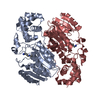
Mass: 55.845 Da / Num. of mol.: 1 / Source method: obtained synthetically / Formula: Fe
#3: Chemical ChemComp-SPD / SPERMIDINE / N-(2-AMINO-PROPYL)-1,4-DIAMINOBUTANE / PA(34)


Mass: 145.246 Da / Num. of mol.: 2 / Source method: obtained synthetically / Formula: C7H19N3
#4: Water ChemComp-HOH / water


Mass: 18.015 Da / Num. of mol.: 164 / Source method: isolated from a natural source / Formula: H2O
Sequence detailsIn chain A, residue numbers 344, 345 are simply skipped. In chain H, residue numbers 345-347 are ...In chain A, residue numbers 344, 345 are simply skipped. In chain H, residue numbers 345-347 are simply skipped.

-
Experimental details

-
Experiment

ExperimentMethod: X-RAY DIFFRACTION / Number of used crystals: 1

-
Sample preparation

CrystalDensity Matthews: 1.95 Å3/Da / Density % sol: 36.86 %
Crystal growTemperature: 293 K / Method: vapor diffusion, sitting drop / pH: 8.5
Details: 0.1M Sodium chloride, 0.1M Tris pH 8.5, 12 % w/v PEG 4000

-
Data collection

DiffractionMean temperature: 100 K
Diffraction sourceSource: SYNCHROTRON / Site: SPring-8 / Beamline: BL44XU / Wavelength: 0.9 Å
DetectorType: RAYONIX MX300HE / Detector: CCD / Date: Dec 13, 2014
RadiationProtocol: SINGLE WAVELENGTH / Monochromatic (M) / Laue (L): M / Scattering type: x-ray
Radiation wavelengthWavelength: 0.9 Å / Relative weight: 1
ReflectionResolution: 1.95→50.01 Å / Num. obs: 47518 / % possible obs: 99.9 % / Redundancy: 5.6 % / Net I/σ(I): 12.4

-
Processing

Software
NameVersionClassification
REFMAC5.8.0151refinement
HKL-2000data reduction
HKL-2000data scaling
MOLREPphasing
RefinementResolution: 1.95→50.01 Å / Cor.coef. Fo:Fc: 0.953 / Cor.coef. Fo:Fc free: 0.93 / SU B: 4.439 / SU ML: 0.126 / Cross valid method: THROUGHOUT / ESU R: 0.215 / ESU R Free: 0.181 / Details: HYDROGENS HAVE BEEN ADDED IN THE RIDING POSITIONS
RfactorNum. reflection% reflectionSelection details
Rfree0.22493 1964 4.9 %RANDOM
Rwork0.17122 ---
obs0.17391 38437 84.8 %-
Solvent computationIon probe radii: 0.8 Å / Shrinkage radii: 0.8 Å / VDW probe radii: 1.2 Å
Displacement parametersBiso mean: 25.426 Å2
Baniso -1Baniso -2Baniso -3
1-1.41 Å2-0 Å20.24 Å2
2---0.53 Å20 Å2
3----0.87 Å2
Refinement stepCycle: 1 / Resolution: 1.95→50.01 Å
ProteinNucleic acidLigandSolventTotal
Num. atoms5478 0 21 164 5663
Refine LS restraints
Refine-IDTypeDev idealDev ideal targetNumber
X-RAY DIFFRACTIONr_bond_refined_d0.0170.0195615
X-RAY DIFFRACTIONr_bond_other_d0.0020.025379
X-RAY DIFFRACTIONr_angle_refined_deg1.8041.9647600
X-RAY DIFFRACTIONr_angle_other_deg1.094312372
X-RAY DIFFRACTIONr_dihedral_angle_1_deg6.6755674
X-RAY DIFFRACTIONr_dihedral_angle_2_deg37.01223.636275
X-RAY DIFFRACTIONr_dihedral_angle_3_deg16.46715980
X-RAY DIFFRACTIONr_dihedral_angle_4_deg18.5831548
X-RAY DIFFRACTIONr_chiral_restr0.1110.2850
X-RAY DIFFRACTIONr_gen_planes_refined0.0080.0216258
X-RAY DIFFRACTIONr_gen_planes_other0.0020.021286
X-RAY DIFFRACTIONr_nbd_refined
X-RAY DIFFRACTIONr_nbd_other
X-RAY DIFFRACTIONr_nbtor_refined
X-RAY DIFFRACTIONr_nbtor_other
X-RAY DIFFRACTIONr_xyhbond_nbd_refined
X-RAY DIFFRACTIONr_xyhbond_nbd_other
X-RAY DIFFRACTIONr_metal_ion_refined
X-RAY DIFFRACTIONr_metal_ion_other
X-RAY DIFFRACTIONr_symmetry_vdw_refined
X-RAY DIFFRACTIONr_symmetry_vdw_other
X-RAY DIFFRACTIONr_symmetry_hbond_refined
X-RAY DIFFRACTIONr_symmetry_hbond_other
X-RAY DIFFRACTIONr_symmetry_metal_ion_refined
X-RAY DIFFRACTIONr_symmetry_metal_ion_other
X-RAY DIFFRACTIONr_mcbond_it2.0312.2922708
X-RAY DIFFRACTIONr_mcbond_other2.0292.2922707
X-RAY DIFFRACTIONr_mcangle_it2.9983.4193375
X-RAY DIFFRACTIONr_mcangle_other2.9983.423376
X-RAY DIFFRACTIONr_scbond_it2.9622.7542907
X-RAY DIFFRACTIONr_scbond_other2.9612.7542908
X-RAY DIFFRACTIONr_scangle_it
X-RAY DIFFRACTIONr_scangle_other4.7763.9774225
X-RAY DIFFRACTIONr_long_range_B_refined7.20344.03323823
X-RAY DIFFRACTIONr_long_range_B_other7.19844.04123728
X-RAY DIFFRACTIONr_rigid_bond_restr
X-RAY DIFFRACTIONr_sphericity_free
X-RAY DIFFRACTIONr_sphericity_bonded
LS refinement shellResolution: 1.949→2 Å / Total num. of bins used: 20
RfactorNum. reflection% reflection
Rfree0.234 108 -
Rwork0.202 1530 -
obs--46.92 %

+
About Yorodumi

-
News

-
Feb 9, 2022. New format data for meta-information of EMDB entries

New format data for meta-information of EMDB entries

  • Version 3 of the EMDB header file is now the official format.
  • The previous official version 1.9 will be removed from the archive.

Related info.:EMDB header

External links:wwPDB to switch to version 3 of the EMDB data model

-
Aug 12, 2020. Covid-19 info

Covid-19 info

URL: https://pdbjlvh1.pdbj.org/emnavi/covid19.php

New page: Covid-19 featured information page in EM Navigator.

Related info.:Covid-19 info / Mar 5, 2020. Novel coronavirus structure data

+
Mar 5, 2020. Novel coronavirus structure data

Novel coronavirus structure data

Related info.:Yorodumi Speices / Aug 12, 2020. Covid-19 info

External links:COVID-19 featured content - PDBj / Molecule of the Month (242):Coronavirus Proteases

+
Jan 31, 2019. EMDB accession codes are about to change! (news from PDBe EMDB page)

EMDB accession codes are about to change! (news from PDBe EMDB page)

  • The allocation of 4 digits for EMDB accession codes will soon come to an end. Whilst these codes will remain in use, new EMDB accession codes will include an additional digit and will expand incrementally as the available range of codes is exhausted. The current 4-digit format prefixed with “EMD-” (i.e. EMD-XXXX) will advance to a 5-digit format (i.e. EMD-XXXXX), and so on. It is currently estimated that the 4-digit codes will be depleted around Spring 2019, at which point the 5-digit format will come into force.
  • The EM Navigator/Yorodumi systems omit the EMD- prefix.

Related info.:Q: What is EMD? / ID/Accession-code notation in Yorodumi/EM Navigator

External links:EMDB Accession Codes are Changing Soon! / Contact to PDBj

+
Jul 12, 2017. Major update of PDB

Major update of PDB

  • wwPDB released updated PDB data conforming to the new PDBx/mmCIF dictionary.
  • This is a major update changing the version number from 4 to 5, and with Remediation, in which all the entries are updated.
  • In this update, many items about electron microscopy experimental information are reorganized (e.g. em_software).
  • Now, EM Navigator and Yorodumi are based on the updated data.

External links:wwPDB Remediation / Enriched Model Files Conforming to OneDep Data Standards Now Available in the PDB FTP Archive

-
Yorodumi

Thousand views of thousand structures

  • Yorodumi is a browser for structure data from EMDB, PDB, SASBDB, etc.
  • This page is also the successor to EM Navigator detail page, and also detail information page/front-end page for Omokage search.
  • The word "yorodu" (or yorozu) is an old Japanese word meaning "ten thousand". "mi" (miru) is to see.

Related info.:EMDB / PDB / SASBDB / Comparison of 3 databanks / Yorodumi Search / Aug 31, 2016. New EM Navigator & Yorodumi / Yorodumi Papers / Jmol/JSmol / Function and homology information / Changes in new EM Navigator and Yorodumi

Read more