 Movie
Movie Controller

[English] 日本語
 Yorodumi
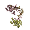
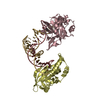
- PDB-5xma: Crystal structure of AsfvPolX in complex with DNA enzyme at P4321... -
+ Open data

- Basic information
| Entry | Database: PDB / ID: 5xma | ||||||
|---|---|---|---|---|---|---|---|
| Title | Crystal structure of AsfvPolX in complex with DNA enzyme at P43212 space group | ||||||
 Components Components |
| ||||||
 Keywords Keywords | TRANSFERASE/DNA / PolX / DNA enzyme / complex / TRANSFERASE-DNA complex | ||||||
| Function / homology |  Function and homology information Function and homology informationvirion component / double-strand break repair via nonhomologous end joining / DNA-directed DNA polymerase / DNA-directed DNA polymerase activity / DNA binding / metal ion binding Similarity search - Function | ||||||
| Biological species |   African swine fever virus African swine fever virussynthetic construct (others) | ||||||
| Method |  X-RAY DIFFRACTION / SYNCHROTRON / MOLECULAR REPLACEMENT / Resolution: 3.8 Å X-RAY DIFFRACTION / SYNCHROTRON / MOLECULAR REPLACEMENT / Resolution: 3.8 Å | ||||||
 Authors Authors | Liu, H.H. / Gan, J.H. | ||||||
| Funding support |  China, 1items China, 1items
| ||||||
 Citation Citation | Journal: Nat Commun / Year: 2017 Title: Crystal structure of an RNA-cleaving DNAzyme. Authors: Liu, H. / Yu, X. / Chen, Y. / Zhang, J. / Wu, B. / Zheng, L. / Haruehanroengra, P. / Wang, R. / Li, S. / Lin, J. / Li, J. / Sheng, J. / Huang, Z. / Ma, J. / Gan, J. | ||||||
| History |
|
- Structure visualization
| Structure viewer | Molecule: MolmilJmol/JSmol |
|---|
- Downloads & links
-Download
| PDBx/mmCIF format | 5xma.cif.gz | 117.1 KB | Display | PDBx/mmCIF format |
|---|---|---|---|---|
| PDB format | pdb5xma.ent.gz | 84.9 KB | Display | PDB format |
| PDBx/mmJSON format | 5xma.json.gz | Tree view | PDBx/mmJSON format | |
| Others |  Other downloads Other downloads |
-Validation report
| Arichive directory | https://data.pdbj.org/pub/pdb/validation_reports/xm/5xmaftp://data.pdbj.org/pub/pdb/validation_reports/xm/5xma | HTTPS FTP |
|---|
-Related structure data
| Related structure data |  5xm8C  5xm9C  5hrbS S: Starting model for refinement C: citing same article ( |
|---|---|
| Similar structure data |


-Links
 PDBj
PDBj


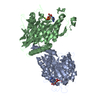
- Assembly
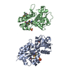
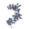
| Deposited unit | 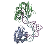
| ||||||||
|---|---|---|---|---|---|---|---|---|---|
| 1 |
| ||||||||
| Unit cell |
|
-Components
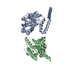
| #1: Protein | Mass: 20552.668 Da / Num. of mol.: 2 Source method: isolated from a genetically manipulated source Source: (gene. exp.) African swine fever virus (strain Badajoz 1971 Vero-adapted)Strain: Badajoz 1971 Vero-adapted / Gene: Ba71V-97, O174L / Production host:  #2: DNA chain | | Mass: 10989.054 Da / Num. of mol.: 1 / Source method: obtained synthetically / Source: (synth.) synthetic construct (others) #3: DNA/RNA hybrid | | Mass: 7210.659 Da / Num. of mol.: 1 / Source method: obtained synthetically / Source: (synth.) synthetic construct (others) |
|---|
-Experimental details
-Experiment
| Experiment | Method: X-RAY DIFFRACTION / Number of used crystals: 1 |
|---|
- Sample preparation
| Crystal | Density Matthews: 3.17 Å3/Da / Density % sol: 61.16 % / Description: diamond |
|---|---|
| Crystal grow | Temperature: 293 K / Method: vapor diffusion, hanging drop / pH: 7 / Details: 0.1 M Tris pH 7.0, 15% v/v ethanol |
-Data collection
| Diffraction | Mean temperature: 100 K |
|---|---|
| Diffraction source | Source: SYNCHROTRON / Site: SSRF / Beamline: BL19U1 / Wavelength: 0.979 Å |
| Detector | Type: DECTRIS PILATUS3 S 6M / Detector: PIXEL / Date: Jun 30, 2016 |
| Radiation | Protocol: SINGLE WAVELENGTH / Monochromatic (M) / Laue (L): M / Scattering type: x-ray |
| Radiation wavelength | Wavelength: 0.979 Å / Relative weight: 1 |
| Reflection | Resolution: 3.8→30 Å / Num. obs: 7857 / % possible obs: 96.8 % / Redundancy: 8.7 % / Rmerge(I) obs: 0.06 / Net I/σ(I): 12.2 |
| Reflection shell | Resolution: 3.8→3.94 Å / Redundancy: 4.1 % / Rmerge(I) obs: 0.37 / Mean I/σ(I) obs: 1.5 / Num. unique obs: 734 / % possible all: 92.9 |
- Processing
| Software |
| ||||||||||||||||||||||||||||
|---|---|---|---|---|---|---|---|---|---|---|---|---|---|---|---|---|---|---|---|---|---|---|---|---|---|---|---|---|---|
| Refinement | Method to determine structure: MOLECULAR REPLACEMENT Starting model: 5HRB Resolution: 3.8→29.353 Å / SU ML: 0.54 / Cross valid method: FREE R-VALUE / σ(F): 1.47 / Phase error: 33.2
| ||||||||||||||||||||||||||||
| Solvent computation | Shrinkage radii: 0.9 Å / VDW probe radii: 1.11 Å | ||||||||||||||||||||||||||||
| Refinement step | Cycle: LAST / Resolution: 3.8→29.353 Å
| ||||||||||||||||||||||||||||
| Refine LS restraints |
| ||||||||||||||||||||||||||||
| LS refinement shell |
|
