 Movie
Movie Controller


+ Open data

- Basic information
| Entry | Database: PDB / ID: 5xlm | ||||||||||||
|---|---|---|---|---|---|---|---|---|---|---|---|---|---|
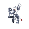
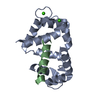
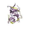
| Title | Monomer form of M.tuberculosis PknI sensor domain | ||||||||||||
 Components Components | Serine/threonine-protein kinase PknI | ||||||||||||
 Keywords Keywords | TRANSFERASE / monomer / sensor domain / PknI / M.tuberculosis | ||||||||||||
| Function / homology |  Function and homology information Function and homology informationregulation of growth rate / peptidyl-threonine phosphorylation / manganese ion binding / peptidyl-serine phosphorylation / protein autophosphorylation / non-specific serine/threonine protein kinase / protein kinase activity / protein serine kinase activity / protein serine/threonine kinase activity / ATP binding ...regulation of growth rate / peptidyl-threonine phosphorylation / manganese ion binding / peptidyl-serine phosphorylation / protein autophosphorylation / non-specific serine/threonine protein kinase / protein kinase activity / protein serine kinase activity / protein serine/threonine kinase activity / ATP binding / plasma membrane / cytosol Similarity search - Function | ||||||||||||
| Biological species |   Mycobacterium tuberculosis (bacteria) Mycobacterium tuberculosis (bacteria) | ||||||||||||
| Method |  X-RAY DIFFRACTION / SYNCHROTRON / MOLECULAR REPLACEMENT / Resolution: 2.2 Å X-RAY DIFFRACTION / SYNCHROTRON / MOLECULAR REPLACEMENT / Resolution: 2.2 Å | ||||||||||||
 Authors Authors | Rao, Z. / Yan, Q. | ||||||||||||
| Funding support |  China, 3items China, 3items
| ||||||||||||
 Citation Citation | Journal: Structure / Year: 2017 Title: Structural Insight into the Activation of PknI Kinase from M. tuberculosis via Dimerization of the Extracellular Sensor Domain. Authors: Yan, Q. / Jiang, D. / Qian, L. / Zhang, Q. / Zhang, W. / Zhou, W. / Mi, K. / Guddat, L. / Yang, H. / Rao, Z. | ||||||||||||
| History |
|
- Structure visualization
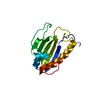
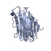
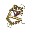
| Structure viewer | Molecule: MolmilJmol/JSmol |
|---|
- Downloads & links
-Download
| PDBx/mmCIF format | 5xlm.cif.gz | 125.6 KB | Display | PDBx/mmCIF format |
|---|---|---|---|---|
| PDB format | pdb5xlm.ent.gz | 101 KB | Display | PDB format |
| PDBx/mmJSON format | 5xlm.json.gz | Tree view | PDBx/mmJSON format | |
| Others |  Other downloads Other downloads |
-Validation report
| Summary document | 5xlm_validation.pdf.gz | 429.8 KB | Display | wwPDB validaton report |
|---|---|---|---|---|
| Full document | 5xlm_full_validation.pdf.gz | 433 KB | Display | |
| Data in XML | 5xlm_validation.xml.gz | 13.3 KB | Display | |
| Data in CIF | 5xlm_validation.cif.gz | 17.5 KB | Display | |
| Arichive directory | https://data.pdbj.org/pub/pdb/validation_reports/xl/5xlmftp://data.pdbj.org/pub/pdb/validation_reports/xl/5xlm | HTTPS FTP |
-Related structure data


-Links
 PDBj
PDBj- Assembly
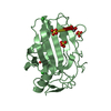
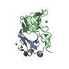
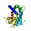
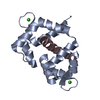
| Deposited unit | 
| ||||||||
|---|---|---|---|---|---|---|---|---|---|
| 1 | 
| ||||||||
| 2 | 
| ||||||||
| Unit cell |
|
-Components
| #1: Protein | Mass: 22630.197 Da / Num. of mol.: 2 / Fragment: UNP RESIDUES 402-585 Source method: isolated from a genetically manipulated source Source: (gene. exp.) Mycobacterium tuberculosis (strain ATCC 25618 / H37Rv) (bacteria)Strain: ATCC 25618 / H37Rv / Gene: pknI, Rv2914c, MTCY338.02c / Production host: References: UniProt: P9WI69, non-specific serine/threonine protein kinase #2: Water | ChemComp-HOH / |  Mass: 18.015 Da / Num. of mol.: 82 / Source method: isolated from a natural source / Formula: H2O Mass: 18.015 Da / Num. of mol.: 82 / Source method: isolated from a natural source / Formula: H2O |
|---|
-Experimental details
-Experiment
| Experiment | Method: X-RAY DIFFRACTION / Number of used crystals: 1 |
|---|
- Sample preparation
| Crystal | Density Matthews: 2.03 Å3/Da / Density % sol: 39.42 % |
|---|---|
| Crystal grow | Temperature: 293 K / Method: vapor diffusion, sitting drop / pH: 7 Details: 1M succinic acid, pH 7.0, 0.1M HEPES, 1% w/v PEG monomethyl ether 2000 |
-Data collection
| Diffraction | Mean temperature: 100 K |
|---|---|
| Diffraction source | Source: SYNCHROTRON / Site: SSRF / Beamline: BL17B1 / Wavelength: 0.97906 Å |
| Detector | Type: ADSC QUANTUM 315 / Detector: CCD / Date: Jul 2, 2013 |
| Radiation | Protocol: SINGLE WAVELENGTH / Monochromatic (M) / Laue (L): M / Scattering type: x-ray |
| Radiation wavelength | Wavelength: 0.97906 Å / Relative weight: 1 |
| Reflection | Resolution: 2.2→50 Å / Num. obs: 18077 / % possible obs: 99.9 % / Redundancy: 5.6 % / Rmerge(I) obs: 0.059 / Rpim(I) all: 0.028 / Net I/σ(I): 28.6 |
| Reflection shell | Resolution: 2.2→2.24 Å / Redundancy: 5.5 % / Rmerge(I) obs: 0.985 / Mean I/σ(I) obs: 1.8 / Num. unique obs: 909 / Rpim(I) all: 0.028 / % possible all: 100 |
- Processing
| Software |
| ||||||||||||||||||||||||||||||||||||||||||
|---|---|---|---|---|---|---|---|---|---|---|---|---|---|---|---|---|---|---|---|---|---|---|---|---|---|---|---|---|---|---|---|---|---|---|---|---|---|---|---|---|---|---|---|
| Refinement | Method to determine structure: MOLECULAR REPLACEMENT / Resolution: 2.2→44.558 Å / SU ML: 0.31 / Cross valid method: FREE R-VALUE / σ(F): 1.98 / Phase error: 31.26
| ||||||||||||||||||||||||||||||||||||||||||
| Solvent computation | Shrinkage radii: 0.9 Å / VDW probe radii: 1.11 Å | ||||||||||||||||||||||||||||||||||||||||||
| Refinement step | Cycle: LAST / Resolution: 2.2→44.558 Å
| ||||||||||||||||||||||||||||||||||||||||||
| Refine LS restraints |
| ||||||||||||||||||||||||||||||||||||||||||
| LS refinement shell |
|
