 Movie
Movie Controller

[English] 日本語
 Yorodumi
- PDB-5xk7: Crystal structure of Isosesquilavandulyl Diphosphate Synthase fro... -
+ Open data

- Basic information
| Entry | Database: PDB / ID: 5xk7 | ||||||
|---|---|---|---|---|---|---|---|
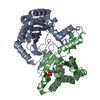
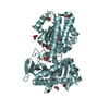
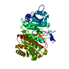
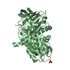
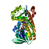
| Title | Crystal structure of Isosesquilavandulyl Diphosphate Synthase from Streptomyces sp. strain CNH-189 in complex with DMAPP | ||||||
 Components Components | Undecaprenyl diphosphate synthase | ||||||
 Keywords Keywords | TRANSFERASE / prenyltransferase / antibiotic biosynthesis | ||||||
| Function / homology | ditrans,polycis-polyprenyl diphosphate synthase [(2E,6E)-farnesyl diphosphate specific] activity / Decaprenyl diphosphate synthase-like / Putative undecaprenyl diphosphate synthase / polyprenol biosynthetic process / Decaprenyl diphosphate synthase-like superfamily / metal ion binding / DIMETHYLALLYL DIPHOSPHATE / PYROPHOSPHATE 2- / Undecaprenyl diphosphate synthase Function and homology information Function and homology information | ||||||
| Biological species |  Streptomyces sp. CNH189 (bacteria) Streptomyces sp. CNH189 (bacteria) | ||||||
| Method |  X-RAY DIFFRACTION / SYNCHROTRON / MOLECULAR REPLACEMENT / Resolution: 1.911 Å X-RAY DIFFRACTION / SYNCHROTRON / MOLECULAR REPLACEMENT / Resolution: 1.911 Å | ||||||
 Authors Authors | Ko, T.P. / Guo, R.T. / Liu, W. / Chen, C.C. / Gao, J. | ||||||
 Citation Citation | Journal: Angew. Chem. Int. Ed. Engl. / Year: 2018 Title: "Head-to-Middle" and "Head-to-Tail" cis-Prenyl Transferases: Structure of Isosesquilavandulyl Diphosphate Synthase. Authors: Gao, J. / Ko, T.P. / Chen, L. / Malwal, S.R. / Zhang, J. / Hu, X. / Qu, F. / Liu, W. / Huang, J.W. / Cheng, Y.S. / Chen, C.C. / Yang, Y. / Zhang, Y. / Oldfield, E. / Guo, R.T. | ||||||
| History |
|
- Structure visualization
| Structure viewer | Molecule: MolmilJmol/JSmol |
|---|
- Downloads & links
-Download
| PDBx/mmCIF format | 5xk7.cif.gz | 397.6 KB | Display | PDBx/mmCIF format |
|---|---|---|---|---|
| PDB format | pdb5xk7.ent.gz | 326.5 KB | Display | PDB format |
| PDBx/mmJSON format | 5xk7.json.gz | Tree view | PDBx/mmJSON format | |
| Others |  Other downloads Other downloads |
-Validation report
| Arichive directory | https://data.pdbj.org/pub/pdb/validation_reports/xk/5xk7ftp://data.pdbj.org/pub/pdb/validation_reports/xk/5xk7 | HTTPS FTP |
|---|
-Related structure data
| Related structure data | 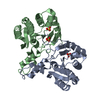 5xk3SC 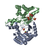 5xk6C  5xk8C 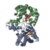 5xk9C S: Starting model for refinement C: citing same article ( |
|---|---|
| Similar structure data |


-Links
 PDBj
PDBj


- Assembly
| Deposited unit | 
| ||||||||
|---|---|---|---|---|---|---|---|---|---|
| 1 | 
| ||||||||
| 2 | 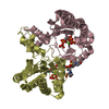
| ||||||||
| Unit cell |
|
-Components
-Protein , 1 types, 4 molecules ABCD
| #1: Protein | Mass: 26708.215 Da / Num. of mol.: 4 Source method: isolated from a genetically manipulated source Source: (gene. exp.) Streptomyces sp. CNH189 (bacteria) / Gene: mcl22 / Plasmid: pET46Ek/LICProduction host: References: UniProt: M4T4U9 |
|---|
-Non-polymers , 6 types, 922 molecules 


| #2: Chemical | ChemComp-MG /  Mass: 24.305 Da / Num. of mol.: 4 / Source method: obtained synthetically / Formula: Mg Mass: 24.305 Da / Num. of mol.: 4 / Source method: obtained synthetically / Formula: Mg#3: Chemical | ChemComp-POP /  Mass: 175.959 Da / Num. of mol.: 5 / Source method: obtained synthetically / Formula: H2O7P2 Mass: 175.959 Da / Num. of mol.: 5 / Source method: obtained synthetically / Formula: H2O7P2#4: Chemical | ChemComp-MES /  Mass: 195.237 Da / Num. of mol.: 4 / Source method: obtained synthetically / Formula: C6H13NO4S / Comment: pH buffer*YM Mass: 195.237 Da / Num. of mol.: 4 / Source method: obtained synthetically / Formula: C6H13NO4S / Comment: pH buffer*YM#5: Chemical |  Mass: 96.063 Da / Num. of mol.: 2 / Source method: obtained synthetically / Formula: SO4 Mass: 96.063 Da / Num. of mol.: 2 / Source method: obtained synthetically / Formula: SO4#6: Chemical |  Mass: 246.092 Da / Num. of mol.: 3 / Source method: obtained synthetically / Formula: C5H12O7P2 Mass: 246.092 Da / Num. of mol.: 3 / Source method: obtained synthetically / Formula: C5H12O7P2#7: Water | ChemComp-HOH / | Mass: 18.015 Da / Num. of mol.: 904 / Source method: isolated from a natural source / Formula: H2O |
|---|
-Experimental details
-Experiment
| Experiment | Method: X-RAY DIFFRACTION / Number of used crystals: 1 |
|---|
- Sample preparation
| Crystal | Density Matthews: 2.42 Å3/Da / Density % sol: 49.23 % |
|---|---|
| Crystal grow | Temperature: 298 K / Method: vapor diffusion, sitting drop Details: PROTEIN: 10 mg/mL in 25 mM Tris-Cl (pH 7.5) and 150 mM NaCl RESERVOIR: 0.2 M (NH4)2SO4, 0.1 M MES pH 6.5, 30% PEG 5000MME SOAK: 5 mM MgCl2, 5 mM (NH4)4P2O7, 0.1 M MES pH 6.5, 30% PEG 5000MME, 10 mM DMAPP |
-Data collection
| Diffraction | Mean temperature: 100 K |
|---|---|
| Diffraction source | Source: SYNCHROTRON / Site: NSRRC  / Beamline: BL15A1 / Wavelength: 1 Å / Beamline: BL15A1 / Wavelength: 1 Å |
| Detector | Type: RAYONIX MX-300 / Detector: CCD / Date: Feb 17, 2017 |
| Radiation | Monochromator: GRAPHITE / Protocol: SINGLE WAVELENGTH / Monochromatic (M) / Laue (L): M / Scattering type: x-ray |
| Radiation wavelength | Wavelength: 1 Å / Relative weight: 1 |
| Reflection | Resolution: 1.91→25 Å / Num. obs: 79758 / % possible obs: 98.5 % / Redundancy: 3.2 % / CC1/2: 0.998 / Rmerge(I) obs: 0.048 / Net I/σ(I): 27.8 |
| Reflection shell | Resolution: 1.91→1.98 Å / Redundancy: 3.3 % / Rmerge(I) obs: 0.459 / Mean I/σ(I) obs: 2.7 / Num. unique obs: 7741 / CC1/2: 0.958 / % possible all: 97.1 |
- Processing
| Software |
| |||||||||||||||||||||||||||||||||||||||||||||||||||||||||||||||||||||||||||||||||||||||||||||||||||||||||
|---|---|---|---|---|---|---|---|---|---|---|---|---|---|---|---|---|---|---|---|---|---|---|---|---|---|---|---|---|---|---|---|---|---|---|---|---|---|---|---|---|---|---|---|---|---|---|---|---|---|---|---|---|---|---|---|---|---|---|---|---|---|---|---|---|---|---|---|---|---|---|---|---|---|---|---|---|---|---|---|---|---|---|---|---|---|---|---|---|---|---|---|---|---|---|---|---|---|---|---|---|---|---|---|---|---|---|
| Refinement | Method to determine structure: MOLECULAR REPLACEMENT Starting model: 5XK3 Resolution: 1.911→24.729 Å / SU ML: 0.19 / Cross valid method: THROUGHOUT / σ(F): 1.35 / Phase error: 20.97
| |||||||||||||||||||||||||||||||||||||||||||||||||||||||||||||||||||||||||||||||||||||||||||||||||||||||||
| Solvent computation | Shrinkage radii: 0.9 Å / VDW probe radii: 1.11 Å | |||||||||||||||||||||||||||||||||||||||||||||||||||||||||||||||||||||||||||||||||||||||||||||||||||||||||
| Refinement step | Cycle: LAST / Resolution: 1.911→24.729 Å
| |||||||||||||||||||||||||||||||||||||||||||||||||||||||||||||||||||||||||||||||||||||||||||||||||||||||||
| Refine LS restraints |
| |||||||||||||||||||||||||||||||||||||||||||||||||||||||||||||||||||||||||||||||||||||||||||||||||||||||||
| LS refinement shell |
| |||||||||||||||||||||||||||||||||||||||||||||||||||||||||||||||||||||||||||||||||||||||||||||||||||||||||
| Refinement TLS params. | Method: refined / Origin x: 7.9075 Å / Origin y: 73.7212 Å / Origin z: 44.5425 Å
| |||||||||||||||||||||||||||||||||||||||||||||||||||||||||||||||||||||||||||||||||||||||||||||||||||||||||
| Refinement TLS group | Selection details: all |
