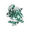
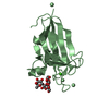
Entry Database : PDB / ID : 5xjvTitle Two intermediate states of conformation switch in dual specificity phosphatase 13a Dual specificity protein phosphatase 13 isoform A Keywords / / / / Function / homology Function Domain/homology Component
/ / / / / / / / / / / / / / / / / / / / / / / / / / / / / / / / Biological species Homo sapiens (human)Method / / / Resolution : 1.69 Å Authors Wei, C.H. / Min, H.G. / Chun, H.J. / Ryu, S.E. Journal : Pharmacol. Res. / Year : 2018Title : Two intermediate states of the conformational switch in dual specificity phosphatase 13aAuthors : Wei, C.H. / Min, H.G. / Kim, M. / Kim, G.H. / Chun, H.J. / Ryu, S.E. History Deposition May 4, 2017 Deposition site / Processing site Revision 1.0 Apr 11, 2018 Provider / Type Revision 1.1 Oct 13, 2021 Group / Database references / Category / diffrn_sourceItem _database_2.pdbx_DOI / _database_2.pdbx_database_accession ... _database_2.pdbx_DOI / _database_2.pdbx_database_accession / _diffrn_source.pdbx_synchrotron_beamline / _diffrn_source.pdbx_synchrotron_site / _diffrn_source.source / _diffrn_source.type Revision 1.2 Nov 22, 2023 Group / Refinement descriptionCategory / chem_comp_bond / pdbx_initial_refinement_model
Show all Show less
 Movie
Movie Controller

 Yorodumi
 Open data
 Basic information
 Components
 Keywords
 Function and homology information
 Homo sapiens (human)
 X-RAY DIFFRACTION /
  Authors
 Citation
 Structure visualization
 Downloads & links
 Other downloads


 PDBj
PDBj


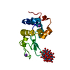
 Assembly


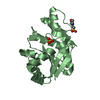
 Mass: 94.971 Da / Num. of mol.: 2 / Source method: obtained synthetically / Formula: PO4
 Mass: 18.015 Da / Num. of mol.: 414 / Source method: isolated from a natural source / Formula: H2O
 Sample preparation
 / Beamline: 7A (6B, 6C1) / Wavelength: 0.97933 Å
 Processing