 Movie
Movie Controller

[English] 日本語
 Yorodumi
- PDB-5xju: Crystal Structure of the Gemin2-binding domain of SMN, Gemin2dN39... -
+ Open data

- Basic information
| Entry | Database: PDB / ID: 5xju | |||||||||
|---|---|---|---|---|---|---|---|---|---|---|
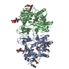
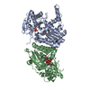
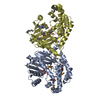
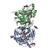
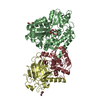
| Title | Crystal Structure of the Gemin2-binding domain of SMN, Gemin2dN39 in Complex with SmD1(1-82)/D2.R61A/F/E/G from Human | |||||||||
 Components Components |
| |||||||||
 Keywords Keywords | SPLICING | |||||||||
| Function / homology |  Function and homology information Function and homology informationGemini of Cajal bodies / SMN complex / U7 snRNP / SLBP independent Processing of Histone Pre-mRNAs / SLBP Dependent Processing of Replication-Dependent Histone Pre-mRNAs / 7-methylguanosine cap hypermethylation / U12-type spliceosomal complex / U1 snRNP binding / pICln-Sm protein complex / methylosome ...Gemini of Cajal bodies / SMN complex / U7 snRNP / SLBP independent Processing of Histone Pre-mRNAs / SLBP Dependent Processing of Replication-Dependent Histone Pre-mRNAs / 7-methylguanosine cap hypermethylation / U12-type spliceosomal complex / U1 snRNP binding / pICln-Sm protein complex / methylosome / RNA splicing, via transesterification reactions / small nuclear ribonucleoprotein complex / SMN-Sm protein complex / spliceosomal tri-snRNP complex / P granule / commitment complex / U2-type precatalytic spliceosome / U2-type prespliceosome assembly / U2-type catalytic step 2 spliceosome / U2-type spliceosomal complex / telomerase holoenzyme complex / U1 snRNP / U2 snRNP / RNA Polymerase II Transcription Termination / U4 snRNP / U2-type prespliceosome / precatalytic spliceosome / mRNA Splicing - Minor Pathway / spliceosomal complex assembly / U5 snRNP / spliceosomal snRNP assembly / Cajal body / U4/U6 x U5 tri-snRNP complex / catalytic step 2 spliceosome / mRNA Splicing - Major Pathway / RNA splicing / spliceosomal complex / mRNA splicing, via spliceosome / DNA-templated transcription termination / Z disc / mRNA processing / cytoplasmic ribonucleoprotein granule / nervous system development / protein-containing complex assembly / snRNP Assembly / SARS-CoV-2 modulates host translation machinery / perikaryon / neuron projection / nuclear body / axon / nucleolus / RNA binding / extracellular exosome / nucleoplasm / identical protein binding / nucleus / cytoplasm / cytosol Similarity search - Function | |||||||||
| Biological species |  Homo sapiens (human) Homo sapiens (human) | |||||||||
| Method |  X-RAY DIFFRACTION / SYNCHROTRON / MOLECULAR REPLACEMENT / Resolution: 2.58 Å X-RAY DIFFRACTION / SYNCHROTRON / MOLECULAR REPLACEMENT / Resolution: 2.58 Å | |||||||||
 Authors Authors | Yi, H. / Zhang, R. | |||||||||
| Funding support |  China, 2items China, 2items
| |||||||||
 Citation Citation | Journal: To Be Published Title: Structures of 7S mutant complexes. Authors: Yi, H. / Zhang, R. | |||||||||
| History |
|
- Structure visualization
| Structure viewer | Molecule: MolmilJmol/JSmol |
|---|
- Downloads & links
-Download
| PDBx/mmCIF format | 5xju.cif.gz | 139.2 KB | Display | PDBx/mmCIF format |
|---|---|---|---|---|
| PDB format | pdb5xju.ent.gz | 106.7 KB | Display | PDB format |
| PDBx/mmJSON format | 5xju.json.gz | Tree view | PDBx/mmJSON format | |
| Others |  Other downloads Other downloads |
-Validation report
| Arichive directory | https://data.pdbj.org/pub/pdb/validation_reports/xj/5xjuftp://data.pdbj.org/pub/pdb/validation_reports/xj/5xju | HTTPS FTP |
|---|
-Related structure data
| Related structure data |  3s6n S: Starting model for refinement |
|---|---|
| Similar structure data |


-Links
 PDBj
PDBj


- Assembly
| Deposited unit | 
| ||||||||
|---|---|---|---|---|---|---|---|---|---|
| 1 |
| ||||||||
| Unit cell |
|
-Components
-Small nuclear ribonucleoprotein ... , 5 types, 5 molecules ABEFG
| #2: Protein | Mass: 9300.950 Da / Num. of mol.: 1 / Fragment: UNP residues 1-82 Source method: isolated from a genetically manipulated source Source: (gene. exp.) Homo sapiens (human) / Gene: SNRPD1 / Production host:  |
|---|---|
| #3: Protein | Mass: 13465.812 Da / Num. of mol.: 1 / Mutation: R61A Source method: isolated from a genetically manipulated source Source: (gene. exp.) Homo sapiens (human) / Gene: SNRPD2, SNRPD1 / Production host: |
| #4: Protein | Mass: 10817.601 Da / Num. of mol.: 1 Source method: isolated from a genetically manipulated source Source: (gene. exp.) Homo sapiens (human) / Gene: SNRPE / Production host: |
| #5: Protein | Mass: 9734.171 Da / Num. of mol.: 1 Source method: isolated from a genetically manipulated source Source: (gene. exp.) Homo sapiens (human) / Gene: SNRPF, PBSCF / Production host: |
| #6: Protein | Mass: 8508.084 Da / Num. of mol.: 1 Source method: isolated from a genetically manipulated source Source: (gene. exp.) Homo sapiens (human) / Gene: SNRPG, PBSCG / Production host: |
-Protein / Protein/peptide , 2 types, 2 molecules 2M
| #1: Protein | Mass: 27323.041 Da / Num. of mol.: 1 / Fragment: UNP residues 40-280 Source method: isolated from a genetically manipulated source Source: (gene. exp.) Homo sapiens (human) / Gene: GEMIN2, SIP1 / Production host: |
|---|---|
| #7: Protein/peptide | Mass: 4047.395 Da / Num. of mol.: 1 / Fragment: UNP residues 26-62 Source method: isolated from a genetically manipulated source Source: (gene. exp.) Homo sapiens (human) / Gene: SMN1, SMN, SMNT, SMN2, SMNC / Production host: |
-Experimental details
-Experiment
| Experiment | Method: X-RAY DIFFRACTION / Number of used crystals: 1 |
|---|
- Sample preparation
| Crystal | Density Matthews: 3.76 Å3/Da / Density % sol: 67.27 % |
|---|---|
| Crystal grow | Temperature: 293 K / Method: vapor diffusion, hanging drop / Details: 1% PEG8000, 100mM Tris.HCl / PH range: 7.5-8.2 |
-Data collection
| Diffraction | Mean temperature: 100 K |
|---|---|
| Diffraction source | Source: SYNCHROTRON / Site: SSRF / Beamline: BL19U1 / Wavelength: 0.97853 Å |
| Detector | Type: MARMOSAIC 225 mm CCD / Detector: CCD / Date: Dec 14, 2015 |
| Radiation | Protocol: SINGLE WAVELENGTH / Monochromatic (M) / Laue (L): M / Scattering type: x-ray |
| Radiation wavelength | Wavelength: 0.97853 Å / Relative weight: 1 |
| Reflection | Resolution: 2.58→66.77 Å / Num. obs: 31276 / % possible obs: 99.9 % / Redundancy: 12.9 % / Net I/σ(I): 13.5 |
- Processing
| Software |
| ||||||||||||||||||||||||||||||||||||||||||||||||||||||||||||
|---|---|---|---|---|---|---|---|---|---|---|---|---|---|---|---|---|---|---|---|---|---|---|---|---|---|---|---|---|---|---|---|---|---|---|---|---|---|---|---|---|---|---|---|---|---|---|---|---|---|---|---|---|---|---|---|---|---|---|---|---|---|
| Refinement | Method to determine structure: MOLECULAR REPLACEMENT Starting model: 3S6N 3s6n Resolution: 2.58→66.77 Å / Cor.coef. Fo:Fc: 0.923 / Cor.coef. Fo:Fc free: 0.879 / SU B: 9.704 / SU ML: 0.206 / Cross valid method: THROUGHOUT / σ(F): 0 / ESU R: 0.547 / ESU R Free: 0.334 / Stereochemistry target values: MAXIMUM LIKELIHOOD Details: HYDROGENS HAVE BEEN ADDED IN THE RIDING POSITIONS U VALUES : REFINED INDIVIDUALLY
| ||||||||||||||||||||||||||||||||||||||||||||||||||||||||||||
| Solvent computation | Ion probe radii: 0.8 Å / Shrinkage radii: 0.8 Å / VDW probe radii: 1.2 Å / Solvent model: MASK | ||||||||||||||||||||||||||||||||||||||||||||||||||||||||||||
| Displacement parameters | Biso max: 185.51 Å2 / Biso mean: 54.285 Å2 / Biso min: 11.76 Å2
| ||||||||||||||||||||||||||||||||||||||||||||||||||||||||||||
| Refinement step | Cycle: final / Resolution: 2.58→66.77 Å
| ||||||||||||||||||||||||||||||||||||||||||||||||||||||||||||
| Refine LS restraints |
| ||||||||||||||||||||||||||||||||||||||||||||||||||||||||||||
| LS refinement shell | Resolution: 2.585→2.652 Å / Rfactor Rfree error: 0 / Total num. of bins used: 20
|
