 Movie
Movie Controller

[English] 日本語
 Yorodumi
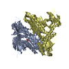
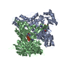
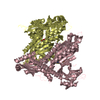
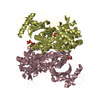
- PDB-5xih: Crystal Structure of Toxoplasma gondii Prolyl-tRNA Synthetase (Tg... -
+ Open data

- Basic information
| Entry | Database: PDB / ID: 5xih | ||||||
|---|---|---|---|---|---|---|---|
| Title | Crystal Structure of Toxoplasma gondii Prolyl-tRNA Synthetase (TgPRS) in complex with inhibitor 5 | ||||||
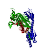
 Components Components | Prolyl-tRNA synthetase (ProRS) | ||||||
 Keywords Keywords | LIGASE / Protein Translation / PRS / Synthetase / Inhibitor / Infectious Disease | ||||||
| Function / homology |  Function and homology information Function and homology informationproline-tRNA ligase / proline-tRNA ligase activity / prolyl-tRNA aminoacylation / aminoacyl-tRNA synthetase multienzyme complex / aminoacyl-tRNA deacylase activity / ATP binding / cytoplasm Similarity search - Function | ||||||
| Biological species |  | ||||||
| Method |  X-RAY DIFFRACTION / SYNCHROTRON / MOLECULAR REPLACEMENT / Resolution: 2.2 Å X-RAY DIFFRACTION / SYNCHROTRON / MOLECULAR REPLACEMENT / Resolution: 2.2 Å | ||||||
 Authors Authors | Jain, V. / Manickam, Y. / Sharma, A. | ||||||
 Citation Citation | Journal: Structure / Year: 2017 Title: Targeting Prolyl-tRNA Synthetase to Accelerate Drug Discovery against Malaria, Leishmaniasis, Toxoplasmosis, Cryptosporidiosis, and Coccidiosis Authors: Jain, V. / Yogavel, M. / Kikuchi, H. / Oshima, Y. / Hariguchi, N. / Matsumoto, M. / Goel, P. / Touquet, B. / Jumani, R.S. / Tacchini-Cottier, F. / Harlos, K. / Huston, C.D. / Hakimi, M.A. / Sharma, A. | ||||||
| History |
|
- Structure visualization
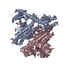
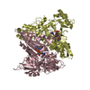
| Structure viewer | Molecule: MolmilJmol/JSmol |
|---|
- Downloads & links
-Download
| PDBx/mmCIF format | 5xih.cif.gz | 415.9 KB | Display | PDBx/mmCIF format |
|---|---|---|---|---|
| PDB format | pdb5xih.ent.gz | 335.9 KB | Display | PDB format |
| PDBx/mmJSON format | 5xih.json.gz | Tree view | PDBx/mmJSON format | |
| Others |  Other downloads Other downloads |
-Validation report
| Arichive directory | https://data.pdbj.org/pub/pdb/validation_reports/xi/5xihftp://data.pdbj.org/pub/pdb/validation_reports/xi/5xih | HTTPS FTP |
|---|
-Related structure data
| Related structure data |  5xifC  5xigC  5xiiC  5xijC  5xikC  5xilC  5xioC  5xipC  5xiqC  4twaS C: citing same article ( S: Starting model for refinement |
|---|---|
| Similar structure data |

-Links
 PDBj
PDBj


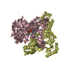
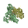
- Assembly
| Deposited unit | 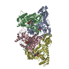
| ||||||||
|---|---|---|---|---|---|---|---|---|---|
| 1 | 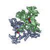
| ||||||||
| 2 | 
| ||||||||
| Unit cell |
|
-Components
-Protein , 1 types, 4 molecules ABCD
| #1: Protein | Mass: 57937.258 Da / Num. of mol.: 4 / Fragment: UNP residues 334-830 Source method: isolated from a genetically manipulated source Source: (gene. exp.) Strain: ATCC 50611 / Me49 / Gene: TGME49_219850 / Plasmid: PETM41 / Production host:  |
|---|
-Non-polymers , 5 types, 676 molecules 

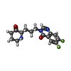


| #2: Chemical | ChemComp-ANP /  Mass: 506.196 Da / Num. of mol.: 4 / Source method: obtained synthetically / Formula: C10H17N6O12P3 / Comment: AMP-PNP, energy-carrying molecule analogue*YM Mass: 506.196 Da / Num. of mol.: 4 / Source method: obtained synthetically / Formula: C10H17N6O12P3 / Comment: AMP-PNP, energy-carrying molecule analogue*YM#3: Chemical | ChemComp-MG /  Mass: 24.305 Da / Num. of mol.: 8 / Source method: obtained synthetically / Formula: Mg Mass: 24.305 Da / Num. of mol.: 8 / Source method: obtained synthetically / Formula: Mg#4: Chemical | ChemComp-86U /  Mass: 321.322 Da / Num. of mol.: 4 / Source method: obtained synthetically / Formula: C16H17F2N3O2 Mass: 321.322 Da / Num. of mol.: 4 / Source method: obtained synthetically / Formula: C16H17F2N3O2#5: Chemical | ChemComp-CL /  Mass: 35.453 Da / Num. of mol.: 6 / Source method: obtained synthetically / Formula: Cl Mass: 35.453 Da / Num. of mol.: 6 / Source method: obtained synthetically / Formula: Cl#6: Water | ChemComp-HOH / | Mass: 18.015 Da / Num. of mol.: 654 / Source method: isolated from a natural source / Formula: H2O |
|---|
-Experimental details
-Experiment
| Experiment | Method: X-RAY DIFFRACTION / Number of used crystals: 1 |
|---|
- Sample preparation
| Crystal | Density Matthews: 2.67 Å3/Da / Density % sol: 53.95 % |
|---|---|
| Crystal grow | Temperature: 293 K / Method: vapor diffusion, hanging drop / pH: 7.5 Details: 10%(w/v) PEG 8K, 20%(v/v) ethylene glycol, 0.03M of each divalent cation and 0.1M MOPS/HEPES-Na |
-Data collection
| Diffraction | Mean temperature: 100 K | ||||||||||||||||||||||||||||||||||||||||||||||||
|---|---|---|---|---|---|---|---|---|---|---|---|---|---|---|---|---|---|---|---|---|---|---|---|---|---|---|---|---|---|---|---|---|---|---|---|---|---|---|---|---|---|---|---|---|---|---|---|---|---|
| Diffraction source | Source: SYNCHROTRON / Site: ESRF  / Beamline: BM14 / Wavelength: 0.9797 Å / Beamline: BM14 / Wavelength: 0.9797 Å | ||||||||||||||||||||||||||||||||||||||||||||||||
| Detector | Type: MARMOSAIC 225 mm CCD / Detector: CCD / Date: Mar 23, 2015 | ||||||||||||||||||||||||||||||||||||||||||||||||
| Radiation | Protocol: SINGLE WAVELENGTH / Monochromatic (M) / Laue (L): M / Scattering type: x-ray | ||||||||||||||||||||||||||||||||||||||||||||||||
| Radiation wavelength | Wavelength: 0.9797 Å / Relative weight: 1 | ||||||||||||||||||||||||||||||||||||||||||||||||
| Reflection | Resolution: 2.2→50 Å / Num. obs: 118866 / % possible obs: 98.5 % / Redundancy: 2.8 % / Rmerge(I) obs: 0.02 / Rpim(I) all: 0.014 / Rrim(I) all: 0.024 / Χ2: 0.524 / Net I/σ(I): 13.5 | ||||||||||||||||||||||||||||||||||||||||||||||||
| Reflection shell | Diffraction-ID: 1 / Redundancy: 2.8 %
|
- Processing
| Software |
| |||||||||||||||||||||||||||||||||||||||||||||
|---|---|---|---|---|---|---|---|---|---|---|---|---|---|---|---|---|---|---|---|---|---|---|---|---|---|---|---|---|---|---|---|---|---|---|---|---|---|---|---|---|---|---|---|---|---|---|
| Refinement | Method to determine structure: MOLECULAR REPLACEMENT Starting model: 4TWA Resolution: 2.2→47.06 Å / Cor.coef. Fo:Fc: 0.953 / Cor.coef. Fo:Fc free: 0.926 / SU B: 4.847 / SU ML: 0.125 / SU R Cruickshank DPI: 0.2356 / Cross valid method: THROUGHOUT / σ(F): 0 / ESU R: 0.236 / ESU R Free: 0.194 / Details: U VALUES : REFINED INDIVIDUALLY
| |||||||||||||||||||||||||||||||||||||||||||||
| Solvent computation | Ion probe radii: 0.8 Å / Shrinkage radii: 0.8 Å / VDW probe radii: 1.2 Å | |||||||||||||||||||||||||||||||||||||||||||||
| Displacement parameters | Biso max: 104.12 Å2 / Biso mean: 31.481 Å2 / Biso min: 8.94 Å2
| |||||||||||||||||||||||||||||||||||||||||||||
| Refinement step | Cycle: final / Resolution: 2.2→47.06 Å
| |||||||||||||||||||||||||||||||||||||||||||||
| Refine LS restraints |
| |||||||||||||||||||||||||||||||||||||||||||||
| LS refinement shell | Resolution: 2.205→2.262 Å / Rfactor Rfree error: 0 / Total num. of bins used: 20
|
