 Movie
Movie Controller

[English] 日本語
 Yorodumi
- PDB-5xca: Crystal structure of GH45 endoglucanase EG27II D137A mutant in co... -
+ Open data

- Basic information
| Entry | Database: PDB / ID: 5xca | |||||||||
|---|---|---|---|---|---|---|---|---|---|---|
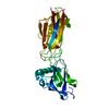
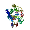
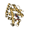
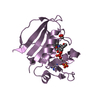
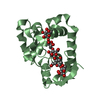
| Title | Crystal structure of GH45 endoglucanase EG27II D137A mutant in complex with cellobiose | |||||||||
 Components Components | Endo-beta-1,4-glucanase | |||||||||
 Keywords Keywords | HYDROLASE / Cellulase / Glycoside Hydrolase Family 45 | |||||||||
| Function / homology |  Function and homology information Function and homology information | |||||||||
| Biological species |  Ampullaria crossean (invertebrata) Ampullaria crossean (invertebrata) | |||||||||
| Method |  X-RAY DIFFRACTION / SYNCHROTRON / MOLECULAR REPLACEMENT / Resolution: 1.35 Å X-RAY DIFFRACTION / SYNCHROTRON / MOLECULAR REPLACEMENT / Resolution: 1.35 Å | |||||||||
 Authors Authors | Nomura, T. / Mizutani, K. / Iwase, H. / Takahashi, N. / Mikami, B. | |||||||||
| Funding support |  Japan, 1items Japan, 1items
| |||||||||
 Citation Citation | Journal: Acta Crystallogr D Struct Biol / Year: 2019 Title: High-resolution crystal structures of the glycoside hydrolase family 45 endoglucanase EG27II from the snail Ampullaria crossean. Authors: Nomura, T. / Iwase, H. / Saka, N. / Takahashi, N. / Mikami, B. / Mizutani, K. | |||||||||
| History |
|
- Structure visualization
| Structure viewer | Molecule: MolmilJmol/JSmol |
|---|
- Downloads & links
-Download
| PDBx/mmCIF format | 5xca.cif.gz | 93.8 KB | Display | PDBx/mmCIF format |
|---|---|---|---|---|
| PDB format | pdb5xca.ent.gz | 70.1 KB | Display | PDB format |
| PDBx/mmJSON format | 5xca.json.gz | Tree view | PDBx/mmJSON format | |
| Others |  Other downloads Other downloads |
-Validation report
| Arichive directory | https://data.pdbj.org/pub/pdb/validation_reports/xc/5xcaftp://data.pdbj.org/pub/pdb/validation_reports/xc/5xca | HTTPS FTP |
|---|
-Related structure data
| Related structure data | 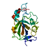 5xbuC 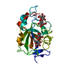 5xbxC 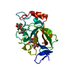 5xc4C 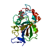 5xc8C 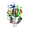 5xc9C  1wc2S C: citing same article ( S: Starting model for refinement |
|---|---|
| Similar structure data |


-Links
 PDBj
PDBj- Assembly
| Deposited unit | 
| ||||||||
|---|---|---|---|---|---|---|---|---|---|
| 1 |
| ||||||||
| Unit cell |
|
-Components
| #1: Protein | Mass: 20871.041 Da / Num. of mol.: 1 / Fragment: UNP residues 17-195 / Mutation: D137A Source method: isolated from a genetically manipulated source Source: (gene. exp.) Ampullaria crossean (invertebrata) / Gene: EG27II / Production host:  Komagataella pastoris (fungus) / Strain (production host): KM71 / References: UniProt: A7KMF0, cellulase Komagataella pastoris (fungus) / Strain (production host): KM71 / References: UniProt: A7KMF0, cellulase |
|---|---|
| #2: Polysaccharide | beta-D-glucopyranose-(1-4)-beta-D-glucopyranose / beta-cellobiose  Source method: isolated from a genetically manipulated source Details: oligosaccharide / References: beta-cellobiose |
| #3: Water | ChemComp-HOH /  Mass: 18.015 Da / Num. of mol.: 175 / Source method: isolated from a natural source / Formula: H2O Mass: 18.015 Da / Num. of mol.: 175 / Source method: isolated from a natural source / Formula: H2O |
| Has protein modification | Y |
-Experimental details
-Experiment
| Experiment | Method: X-RAY DIFFRACTION / Number of used crystals: 1 |
|---|
- Sample preparation
| Crystal | Density Matthews: 1.75 Å3/Da / Density % sol: 29.88 % |
|---|---|
| Crystal grow | Temperature: 293 K / Method: vapor diffusion, hanging drop / pH: 6.5 / Details: 25% PEG4000, 200mM KCl, 50mM MES |
-Data collection
| Diffraction | Mean temperature: 100 K |
|---|---|
| Diffraction source | Source: SYNCHROTRON / Site: SPring-8 / Beamline: BL26B1 / Wavelength: 1 Å |
| Detector | Type: RAYONIX MX225HE / Detector: CCD / Date: Dec 3, 2016 |
| Radiation | Protocol: SINGLE WAVELENGTH / Monochromatic (M) / Laue (L): M / Scattering type: x-ray |
| Radiation wavelength | Wavelength: 1 Å / Relative weight: 1 |
| Reflection | Resolution: 1.35→50 Å / Num. obs: 32869 / % possible obs: 99.5 % / Redundancy: 7 % / Rmerge(I) obs: 0.088 / Net I/σ(I): 33.5 |
| Reflection shell | Resolution: 1.35→1.37 Å / Redundancy: 6.6 % / Rmerge(I) obs: 0.428 / Mean I/σ(I) obs: 4 / % possible all: 99.9 |
- Processing
| Software |
| ||||||||||||||||||||||||||||||||||||||||||||||||||||||||||||||||||||||||||||||||||||
|---|---|---|---|---|---|---|---|---|---|---|---|---|---|---|---|---|---|---|---|---|---|---|---|---|---|---|---|---|---|---|---|---|---|---|---|---|---|---|---|---|---|---|---|---|---|---|---|---|---|---|---|---|---|---|---|---|---|---|---|---|---|---|---|---|---|---|---|---|---|---|---|---|---|---|---|---|---|---|---|---|---|---|---|---|---|
| Refinement | Method to determine structure: MOLECULAR REPLACEMENT Starting model: 1WC2 Resolution: 1.35→41.51 Å / SU ML: 0.15 / Cross valid method: FREE R-VALUE / σ(F): 1.35 / Phase error: 22.65
| ||||||||||||||||||||||||||||||||||||||||||||||||||||||||||||||||||||||||||||||||||||
| Solvent computation | Shrinkage radii: 0.9 Å / VDW probe radii: 1.11 Å | ||||||||||||||||||||||||||||||||||||||||||||||||||||||||||||||||||||||||||||||||||||
| Refinement step | Cycle: LAST / Resolution: 1.35→41.51 Å
| ||||||||||||||||||||||||||||||||||||||||||||||||||||||||||||||||||||||||||||||||||||
| Refine LS restraints |
| ||||||||||||||||||||||||||||||||||||||||||||||||||||||||||||||||||||||||||||||||||||
| LS refinement shell |
|
