 Movie
Movie Controller

[English] 日本語
 Yorodumi
- PDB-5x8s: Crystal Structure of the mutant Human ROR gamma Ligand Binding Do... -
+ Open data

- Basic information
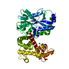
| Entry | Database: PDB / ID: 5x8s | ||||||
|---|---|---|---|---|---|---|---|
| Title | Crystal Structure of the mutant Human ROR gamma Ligand Binding Domain With Ursolic acid. | ||||||
 Components Components | Nuclear receptor ROR-gamma | ||||||
 Keywords Keywords | TRANSFERASE/INHIBITOR / Inhibitor / Binary Complex / Nuclear Receptor / TRANSFERASE-INHIBITOR complex | ||||||
| Function / homology |  Function and homology information Function and homology informationtolerance induction in gut-associated lymphoid tissue / T-helper 17 cell differentiation / cellular response to sterol / regulation of steroid metabolic process / ligand-modulated transcription factor activity / Peyer's patch development / regulatory T cell differentiation / T-helper cell differentiation / positive regulation of circadian rhythm / RUNX3 Regulates Immune Response and Cell Migration ...tolerance induction in gut-associated lymphoid tissue / T-helper 17 cell differentiation / cellular response to sterol / regulation of steroid metabolic process / ligand-modulated transcription factor activity / Peyer's patch development / regulatory T cell differentiation / T-helper cell differentiation / positive regulation of circadian rhythm / RUNX3 Regulates Immune Response and Cell Migration / oxysterol binding / negative regulation of thymocyte apoptotic process / regulation of fat cell differentiation / Phosphorylated BMAL1:CLOCK (ARNTL:CLOCK) activates expression of core clock genes / regulation of glucose metabolic process / lymph node development / adipose tissue development / xenobiotic metabolic process / RORA,B,C and NR1D1 (REV-ERBA) regulate gene expression / Expression of BMAL (ARNTL), CLOCK, and NPAS2 / circadian regulation of gene expression / Nuclear Receptor transcription pathway / DNA-binding transcription repressor activity, RNA polymerase II-specific / nuclear receptor activity / sequence-specific double-stranded DNA binding / Interleukin-4 and Interleukin-13 signaling / DNA-binding transcription factor activity, RNA polymerase II-specific / nuclear body / RNA polymerase II cis-regulatory region sequence-specific DNA binding / DNA-binding transcription factor activity / regulation of transcription by RNA polymerase II / positive regulation of DNA-templated transcription / chromatin / negative regulation of transcription by RNA polymerase II / zinc ion binding / nucleoplasm / nucleus Similarity search - Function | ||||||
| Biological species |  Homo sapiens (human) Homo sapiens (human) | ||||||
| Method |  X-RAY DIFFRACTION / SYNCHROTRON / MOLECULAR REPLACEMENT / Resolution: 2.2 Å X-RAY DIFFRACTION / SYNCHROTRON / MOLECULAR REPLACEMENT / Resolution: 2.2 Å | ||||||
 Authors Authors | Noguchi, M. / Nomura, A. / Murase, K. / Doi, S. / Yamaguchi, K. / Adachi, T. | ||||||
 Citation Citation | Journal: Genes Cells / Year: 2017 Title: Ternary complex of human ROR gamma ligand-binding domain, inverse agonist and SMRT peptide shows a unique mechanism of corepressor recruitment Authors: Noguchi, M. / Nomura, A. / Murase, K. / Doi, S. / Yamaguchi, K. / Hirata, K. / Shiozaki, M. / Hirashima, S. / Kotoku, M. / Yamaguchi, T. / Katsuda, Y. / Steensma, R. / Li, X. / Tao, H. / ...Authors: Noguchi, M. / Nomura, A. / Murase, K. / Doi, S. / Yamaguchi, K. / Hirata, K. / Shiozaki, M. / Hirashima, S. / Kotoku, M. / Yamaguchi, T. / Katsuda, Y. / Steensma, R. / Li, X. / Tao, H. / Tse, B. / Fenn, M. / Babine, R. / Bradley, E. / Crowe, P. / Thacher, S. / Adachi, T. / Kamada, M. | ||||||
| History |
|
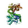
- Structure visualization
| Structure viewer | Molecule: MolmilJmol/JSmol |
|---|
- Downloads & links
-Download
| PDBx/mmCIF format | 5x8s.cif.gz | 116.5 KB | Display | PDBx/mmCIF format |
|---|---|---|---|---|
| PDB format | pdb5x8s.ent.gz | 89.4 KB | Display | PDB format |
| PDBx/mmJSON format | 5x8s.json.gz | Tree view | PDBx/mmJSON format | |
| Others |  Other downloads Other downloads |
-Validation report
| Arichive directory | https://data.pdbj.org/pub/pdb/validation_reports/x8/5x8sftp://data.pdbj.org/pub/pdb/validation_reports/x8/5x8s | HTTPS FTP |
|---|
-Related structure data
| Related structure data |  5x8qC 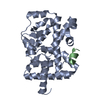 5x8uC 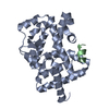 5x8wC  5x8xC 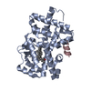 3l0lS C: citing same article ( S: Starting model for refinement |
|---|---|
| Similar structure data |


-Links
 PDBj
PDBj- Assembly
| Deposited unit | 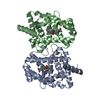
| ||||||||
|---|---|---|---|---|---|---|---|---|---|
| 1 |
| ||||||||
| Unit cell |
|
-Components
| #1: Protein | Mass: 29898.516 Da / Num. of mol.: 2 / Fragment: UNP residues 261-518 Source method: isolated from a genetically manipulated source Source: (gene. exp.) Homo sapiens (human) / Gene: RORC, NR1F3, RORG, RZRG / Production host:  #2: Chemical | 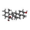  Mass: 456.700 Da / Num. of mol.: 2 / Source method: obtained synthetically / Formula: C30H48O3 Mass: 456.700 Da / Num. of mol.: 2 / Source method: obtained synthetically / Formula: C30H48O3#3: Water | ChemComp-HOH / |  Mass: 18.015 Da / Num. of mol.: 298 / Source method: isolated from a natural source / Formula: H2O Mass: 18.015 Da / Num. of mol.: 298 / Source method: isolated from a natural source / Formula: H2O |
|---|
-Experimental details
-Experiment
| Experiment | Method: X-RAY DIFFRACTION / Number of used crystals: 1 |
|---|
- Sample preparation
| Crystal | Density Matthews: 2.57 Å3/Da / Density % sol: 52.21 % |
|---|---|
| Crystal grow | Temperature: 295 K / Method: vapor diffusion, sitting drop / Details: 0.1M Hepes pH7.5, 5% PEG4000, temperature 295K |
-Data collection
| Diffraction | Mean temperature: 100 K |
|---|---|
| Diffraction source | Source: SYNCHROTRON / Site: Photon Factory  / Beamline: BL-17A / Wavelength: 0.98 Å / Beamline: BL-17A / Wavelength: 0.98 Å |
| Detector | Type: ADSC QUANTUM 315 / Detector: CCD / Date: Nov 8, 2011 |
| Radiation | Protocol: SINGLE WAVELENGTH / Monochromatic (M) / Laue (L): M / Scattering type: x-ray |
| Radiation wavelength | Wavelength: 0.98 Å / Relative weight: 1 |
| Reflection | Resolution: 2.2→101.28 Å / Num. obs: 30947 / % possible obs: 100 % / Redundancy: 21.3 % / Rmerge(I) obs: 0.098 / Net I/σ(I): 11.4 |
| Reflection shell | Resolution: 2.2→2.26 Å / Redundancy: 11.4 % / Rmerge(I) obs: 0.435 / Mean I/σ(I) obs: 1.8 / % possible all: 100 |
- Processing
| Software |
| ||||||||||||||||||||||||||||||||||||||||||||||||||||||||||||||||||||||||||||||||||||||||||||||||||||||||||||||||||||||||||||||||||||||||||||||||||||||||||||||||||||||||||||||||||||||
|---|---|---|---|---|---|---|---|---|---|---|---|---|---|---|---|---|---|---|---|---|---|---|---|---|---|---|---|---|---|---|---|---|---|---|---|---|---|---|---|---|---|---|---|---|---|---|---|---|---|---|---|---|---|---|---|---|---|---|---|---|---|---|---|---|---|---|---|---|---|---|---|---|---|---|---|---|---|---|---|---|---|---|---|---|---|---|---|---|---|---|---|---|---|---|---|---|---|---|---|---|---|---|---|---|---|---|---|---|---|---|---|---|---|---|---|---|---|---|---|---|---|---|---|---|---|---|---|---|---|---|---|---|---|---|---|---|---|---|---|---|---|---|---|---|---|---|---|---|---|---|---|---|---|---|---|---|---|---|---|---|---|---|---|---|---|---|---|---|---|---|---|---|---|---|---|---|---|---|---|---|---|---|---|
| Refinement | Method to determine structure: MOLECULAR REPLACEMENT Starting model: 3L0L Resolution: 2.2→101.28 Å / Cor.coef. Fo:Fc: 0.948 / Cor.coef. Fo:Fc free: 0.899 / SU B: 5.14 / SU ML: 0.132 / Cross valid method: THROUGHOUT / ESU R: 0.228 / ESU R Free: 0.199 / Stereochemistry target values: MAXIMUM LIKELIHOOD
| ||||||||||||||||||||||||||||||||||||||||||||||||||||||||||||||||||||||||||||||||||||||||||||||||||||||||||||||||||||||||||||||||||||||||||||||||||||||||||||||||||||||||||||||||||||||
| Solvent computation | Ion probe radii: 0.8 Å / Shrinkage radii: 0.8 Å / VDW probe radii: 1.2 Å / Solvent model: BABINET MODEL WITH MASK | ||||||||||||||||||||||||||||||||||||||||||||||||||||||||||||||||||||||||||||||||||||||||||||||||||||||||||||||||||||||||||||||||||||||||||||||||||||||||||||||||||||||||||||||||||||||
| Displacement parameters | Biso mean: 30.102 Å2
| ||||||||||||||||||||||||||||||||||||||||||||||||||||||||||||||||||||||||||||||||||||||||||||||||||||||||||||||||||||||||||||||||||||||||||||||||||||||||||||||||||||||||||||||||||||||
| Refinement step | Cycle: 1 / Resolution: 2.2→101.28 Å
| ||||||||||||||||||||||||||||||||||||||||||||||||||||||||||||||||||||||||||||||||||||||||||||||||||||||||||||||||||||||||||||||||||||||||||||||||||||||||||||||||||||||||||||||||||||||
| Refine LS restraints |
|