 Movie
Movie Controller

[English] 日本語
 Yorodumi
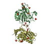
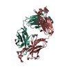
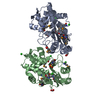
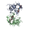
- PDB-5x7v: Crystal structure of Nucleosome assembly protein S (PfNapS) from ... -
+ Open data

- Basic information
| Entry | Database: PDB / ID: 5x7v | |||||||||
|---|---|---|---|---|---|---|---|---|---|---|
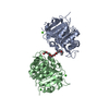
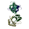
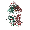
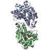
| Title | Crystal structure of Nucleosome assembly protein S (PfNapS) from Plasmodium falciparum | |||||||||
 Components Components | Nucleosome assembly protein | |||||||||
 Keywords Keywords | CHAPERONE / NUCLEOSOME ASSEMBLY PROTEIN / HISTONE RECOGNITION | |||||||||
| Function / homology |  Function and homology information Function and homology information | |||||||||
| Biological species |  | |||||||||
| Method |  X-RAY DIFFRACTION / SYNCHROTRON / SAD / Resolution: 2.802 Å X-RAY DIFFRACTION / SYNCHROTRON / SAD / Resolution: 2.802 Å | |||||||||
 Authors Authors | Gill, J. / Yogavel, M. / Sharma, A. | |||||||||
 Citation Citation | Journal: Malar. J. / Year: 2010 Title: Structure, localization and histone binding properties of nuclear-associated nucleosome assembly protein from Plasmodium falciparum. Authors: Gill, J. / Kumar, A. / Yogavel, M. / Belrhali, H. / Jain, S.K. / Rug, M. / Brown, M. / Maier, A.G. / Sharma, A. | |||||||||
| History |
|
- Structure visualization
| Structure viewer | Molecule: MolmilJmol/JSmol |
|---|
- Downloads & links
-Download
| PDBx/mmCIF format | 5x7v.cif.gz | 212.7 KB | Display | PDBx/mmCIF format |
|---|---|---|---|---|
| PDB format | pdb5x7v.ent.gz | 172 KB | Display | PDB format |
| PDBx/mmJSON format | 5x7v.json.gz | Tree view | PDBx/mmJSON format | |
| Others |  Other downloads Other downloads |
-Validation report
| Arichive directory | https://data.pdbj.org/pub/pdb/validation_reports/x7/5x7vftp://data.pdbj.org/pub/pdb/validation_reports/x7/5x7v | HTTPS FTP |
|---|
-Related structure data
| Similar structure data |
|---|
-Links
 PDBj
PDBj


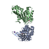
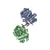
- Assembly
| Deposited unit | 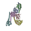
| ||||||||
|---|---|---|---|---|---|---|---|---|---|
| 1 | 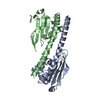
| ||||||||
| 2 | 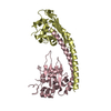
| ||||||||
| 3 | 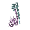
| ||||||||
| Unit cell |
|
-Components
| #1: Protein | Mass: 22993.096 Da / Num. of mol.: 6 / Fragment: UNP RESIDUES 29-221 Source method: isolated from a genetically manipulated source Source: (gene. exp.) Gene: B7 / Production host:  |
|---|
-Experimental details
-Experiment
| Experiment | Method: X-RAY DIFFRACTION / Number of used crystals: 1 |
|---|
- Sample preparation
| Crystal | Density Matthews: 2.77 Å3/Da / Density % sol: 60.09 % |
|---|---|
| Crystal grow | Temperature: 293.15 K / Method: vapor diffusion, hanging drop / pH: 5 Details: 20% PEG 3350, 0.2M di-ammonuium tatrate, VAPOR DIFFUSION, HANGING DROP |
-Data collection
| Diffraction | Mean temperature: 100 K |
|---|---|
| Diffraction source | Source: SYNCHROTRON / Site: ESRF  / Beamline: BM14 / Wavelength: 1 Å / Beamline: BM14 / Wavelength: 1 Å |
| Detector | Type: MARMOSAIC 225 mm CCD / Detector: CCD / Date: Apr 16, 2007 |
| Radiation | Protocol: SINGLE WAVELENGTH / Monochromatic (M) / Laue (L): M / Scattering type: x-ray |
| Radiation wavelength | Wavelength: 1 Å / Relative weight: 1 |
| Reflection | Resolution: 2.8→50 Å / Num. obs: 35024 / % possible obs: 96.6 % / Redundancy: 5.9 % / Net I/σ(I): 23.5 |
| Reflection shell | Resolution: 2.8→2.9 Å / % possible all: 81.8 |
- Processing
| Software |
| |||||||||||||||||||||||||||||||||||||||||||||||||||||||||||||||||||||||||||||||||||||||||||
|---|---|---|---|---|---|---|---|---|---|---|---|---|---|---|---|---|---|---|---|---|---|---|---|---|---|---|---|---|---|---|---|---|---|---|---|---|---|---|---|---|---|---|---|---|---|---|---|---|---|---|---|---|---|---|---|---|---|---|---|---|---|---|---|---|---|---|---|---|---|---|---|---|---|---|---|---|---|---|---|---|---|---|---|---|---|---|---|---|---|---|---|---|
| Refinement | Method to determine structure: SAD / Resolution: 2.802→29.244 Å / SU ML: 0.44 / Cross valid method: FREE R-VALUE / σ(F): 1 / Phase error: 44.2
| |||||||||||||||||||||||||||||||||||||||||||||||||||||||||||||||||||||||||||||||||||||||||||
| Solvent computation | Shrinkage radii: 0.9 Å / VDW probe radii: 1.11 Å | |||||||||||||||||||||||||||||||||||||||||||||||||||||||||||||||||||||||||||||||||||||||||||
| Refinement step | Cycle: LAST / Resolution: 2.802→29.244 Å
| |||||||||||||||||||||||||||||||||||||||||||||||||||||||||||||||||||||||||||||||||||||||||||
| Refine LS restraints |
| |||||||||||||||||||||||||||||||||||||||||||||||||||||||||||||||||||||||||||||||||||||||||||
| LS refinement shell |
|
