[English] 日本語
Yorodumi
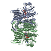
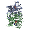
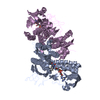
- PDB-5x1y: Structure of mercuric reductase from Lysinibacillus sphaericus -

+
Open data


ID or keywords:

Loading...

-
Basic information

Entry
Database: PDB / ID: 5x1y
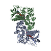
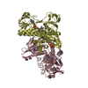
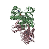
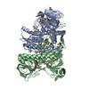
TitleStructure of mercuric reductase from Lysinibacillus sphaericus
ComponentsMercuric reductase
KeywordsOXIDOREDUCTASE / Mercuric reductase / merA / FAD
Function / homology
Function and homology information


mercury(II) reductase / mercury (II) reductase (NADP+) activity / oxidoreductase activity, acting on a sulfur group of donors, NAD(P) as acceptor / detoxification of mercury ion / metal ion transport / mercury ion binding / cell redox homeostasis / NADP binding / flavin adenine dinucleotide binding
Similarity search - Function
Mercury(II) reductase / Heavy-metal-associated domain / Heavy metal-associated domain superfamily / Heavy-metal-associated domain profile. / Heavy metal-associated domain, HMA / Pyridine nucleotide-disulphide oxidoreductase, class I / FAD/NAD-linked reductase, C-terminal dimerisation domain / Pyridine nucleotide-disulphide oxidoreductase, class I, active site / Pyridine nucleotide-disulphide oxidoreductases class-I active site. / Pyridine nucleotide-disulphide oxidoreductase, dimerisation domain ...Mercury(II) reductase / Heavy-metal-associated domain / Heavy metal-associated domain superfamily / Heavy-metal-associated domain profile. / Heavy metal-associated domain, HMA / Pyridine nucleotide-disulphide oxidoreductase, class I / FAD/NAD-linked reductase, C-terminal dimerisation domain / Pyridine nucleotide-disulphide oxidoreductase, class I, active site / Pyridine nucleotide-disulphide oxidoreductases class-I active site. / Pyridine nucleotide-disulphide oxidoreductase, dimerisation domain / Pyridine nucleotide-disulphide oxidoreductase, dimerisation domain / FAD/NAD-linked reductase, dimerisation domain superfamily / FAD/NAD(P)-binding domain / Pyridine nucleotide-disulphide oxidoreductase / Enolase-like; domain 1 / FAD/NAD(P)-binding domain / FAD/NAD(P)-binding domain / 3-Layer(bba) Sandwich / FAD/NAD(P)-binding domain superfamily / 2-Layer Sandwich / Alpha Beta
Similarity search - Domain/homology
FLAVIN-ADENINE DINUCLEOTIDE / Mercuric reductase
Similarity search - Component
Biological speciesLysinibacillus sphaericus (bacteria)
MethodX-RAY DIFFRACTION / SYNCHROTRON / MOLECULAR REPLACEMENT / Resolution: 3.48 Å
AuthorsKhan, F. / Suguna, K.
CitationJournal: Biometals / Year: 2017
Title: Structural and functional characterization of mercuric reductase from Lysinibacillus sphaericus strain G1.
Authors: Bafana, A. / Khan, F. / Suguna, K.
History
DepositionJan 28, 2017Deposition site: PDBJ / Processing site: PDBJ
Revision 1.0Oct 4, 2017Provider: repository / Type: Initial release
Revision 1.1Dec 6, 2017Group: Database references / Category: citation
Item: _citation.journal_volume / _citation.page_first / _citation.page_last
Revision 1.2Nov 22, 2023Group: Data collection / Database references / Refinement description
Category: chem_comp_atom / chem_comp_bond ...chem_comp_atom / chem_comp_bond / citation / database_2 / pdbx_initial_refinement_model
Item: _citation.country / _database_2.pdbx_DOI / _database_2.pdbx_database_accession

-
Structure visualization

Structure viewerMolecule:
MolmilJmol/JSmol

Downloads & links

-
Assembly

Deposited unit
B: Mercuric reductase
A: Mercuric reductase
C: Mercuric reductase
D: Mercuric reductase
E: Mercuric reductase
F: Mercuric reductase
hetero molecules


Theoretical massNumber of molelcules
Total (without water)363,90912
Polymers359,1956
Non-polymers4,7136
Water00
1
B: Mercuric reductase
C: Mercuric reductase
hetero molecules


Theoretical massNumber of molelcules
Total (without water)121,3034
Polymers119,7322
Non-polymers1,5712
Water0
TypeNameSymmetry operationNumber
identity operation1_555x,y,z1
Buried area9490 Å2
ΔGint-69 kcal/mol
Surface area32880 Å2
MethodPISA
2
A: Mercuric reductase
E: Mercuric reductase
hetero molecules


Theoretical massNumber of molelcules
Total (without water)121,3034
Polymers119,7322
Non-polymers1,5712
Water0
TypeNameSymmetry operationNumber
identity operation1_555x,y,z1
Buried area9440 Å2
ΔGint-69 kcal/mol
Surface area33410 Å2
MethodPISA
3
D: Mercuric reductase
F: Mercuric reductase
hetero molecules


Theoretical massNumber of molelcules
Total (without water)121,3034
Polymers119,7322
Non-polymers1,5712
Water0
TypeNameSymmetry operationNumber
identity operation1_555x,y,z1
Buried area9470 Å2
ΔGint-69 kcal/mol
Surface area33460 Å2
MethodPISA
Unit cell
Length a, b, c (Å)236.380, 150.270, 122.780
Angle α, β, γ (deg.)90.00, 92.62, 90.00
Int Tables number5
Space group name H-MC121

-
Components

#1: Protein
Mercuric reductase / Hg(II) reductase


Mass: 59865.875 Da / Num. of mol.: 6
Source method: isolated from a genetically manipulated source
Details: The protein N-terminus got cleaved during crystallization and the electron density for few C-terminus residues is missing .
Source: (gene. exp.) Lysinibacillus sphaericus (bacteria) / Gene: merA / Production host: Escherichia coli (E. coli) / Strain (production host): BL21 / References: UniProt: D9J041, mercury(II) reductase
#2: Chemical
ChemComp-FAD / FLAVIN-ADENINE DINUCLEOTIDE


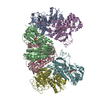
Mass: 785.550 Da / Num. of mol.: 6 / Source method: obtained synthetically / Formula: C27H33N9O15P2 / Comment: FAD*YM

-
Experimental details

-
Experiment

ExperimentMethod: X-RAY DIFFRACTION / Number of used crystals: 1

-
Sample preparation

CrystalDensity Matthews: 3.03 Å3/Da / Density % sol: 59.44 %
Crystal growTemperature: 289 K / Method: microbatch / pH: 7
Details: 5% v/v Tascimate, 0.1 M HEPES, pH 7.0 and 10% w/v PEG monomethyl ether 5000

-
Data collection

DiffractionMean temperature: 100 K
Diffraction sourceSource: SYNCHROTRON / Site: ESRF / Beamline: BM14 / Wavelength: 0.9537 Å
DetectorType: MARMOSAIC 225 mm CCD / Detector: CCD / Date: Apr 30, 2015
RadiationProtocol: SINGLE WAVELENGTH / Monochromatic (M) / Laue (L): M / Scattering type: x-ray
Radiation wavelengthWavelength: 0.9537 Å / Relative weight: 1
ReflectionResolution: 3.48→87.072 Å / Num. obs: 55103 / % possible obs: 100 % / Redundancy: 11 % / Net I/σ(I): 18.93

-
Processing

Software
NameVersionClassification
PHENIX(1.10.1_2155)refinement
iMOSFLMdata reduction
Aimlessdata scaling
PHASERphasing
RefinementMethod to determine structure: MOLECULAR REPLACEMENT
Starting model: 1ZK7
Resolution: 3.48→87.072 Å / SU ML: 0.41 / Cross valid method: FREE R-VALUE / σ(F): 1.34 / Phase error: 22.97 / Stereochemistry target values: ML
RfactorNum. reflection% reflection
Rfree0.2396 2760 5.01 %
Rwork0.1913 --
obs0.1937 55045 99.89 %
Solvent computationShrinkage radii: 0.9 Å / VDW probe radii: 1.11 Å / Solvent model: FLAT BULK SOLVENT MODEL
Refinement stepCycle: LAST / Resolution: 3.48→87.072 Å
ProteinNucleic acidLigandSolventTotal
Num. atoms19777 0 318 0 20095
Refine LS restraints
Refine-IDTypeDev idealNumber
X-RAY DIFFRACTIONf_bond_d0.00320378
X-RAY DIFFRACTIONf_angle_d0.61927765
X-RAY DIFFRACTIONf_dihedral_angle_d12.1712179
X-RAY DIFFRACTIONf_chiral_restr0.0423437
X-RAY DIFFRACTIONf_plane_restr0.0033492
LS refinement shell
Resolution (Å)Rfactor RfreeNum. reflection RfreeRfactor RworkNum. reflection RworkRefine-ID% reflection obs (%)
3.48-3.540.29861110.2432643X-RAY DIFFRACTION100
3.54-3.60440.35121270.24412629X-RAY DIFFRACTION100
3.6044-3.67370.30241120.24992599X-RAY DIFFRACTION100
3.6737-3.74870.26651300.23792620X-RAY DIFFRACTION100
3.7487-3.83020.25511320.2232611X-RAY DIFFRACTION100
3.8302-3.91930.27391360.22672585X-RAY DIFFRACTION100
3.9193-4.01730.25891210.21112640X-RAY DIFFRACTION100
4.0173-4.1260.2731410.20792609X-RAY DIFFRACTION100
4.126-4.24740.28581350.2012637X-RAY DIFFRACTION100
4.2474-4.38450.23591290.17572600X-RAY DIFFRACTION100
4.3845-4.54120.21041340.16892605X-RAY DIFFRACTION100
4.5412-4.7230.20291340.16292611X-RAY DIFFRACTION100
4.723-4.93790.20791810.15492554X-RAY DIFFRACTION100
4.9379-5.19820.20321590.16492593X-RAY DIFFRACTION100
5.1982-5.52380.26991460.18742618X-RAY DIFFRACTION100
5.5238-5.95020.23591650.20532590X-RAY DIFFRACTION100
5.9502-6.54890.26431540.18592612X-RAY DIFFRACTION100
6.5489-7.4960.22041430.17582615X-RAY DIFFRACTION100
7.496-9.44240.17521440.14512651X-RAY DIFFRACTION100
9.4424-87.09950.22191260.18782663X-RAY DIFFRACTION98

+
About Yorodumi

-
News

-
Feb 9, 2022. New format data for meta-information of EMDB entries

New format data for meta-information of EMDB entries

  • Version 3 of the EMDB header file is now the official format.
  • The previous official version 1.9 will be removed from the archive.

Related info.:EMDB header

External links:wwPDB to switch to version 3 of the EMDB data model

-
Aug 12, 2020. Covid-19 info

Covid-19 info

URL: https://pdbjlvh1.pdbj.org/emnavi/covid19.php

New page: Covid-19 featured information page in EM Navigator.

Related info.:Covid-19 info / Mar 5, 2020. Novel coronavirus structure data

+
Mar 5, 2020. Novel coronavirus structure data

Novel coronavirus structure data

Related info.:Yorodumi Speices / Aug 12, 2020. Covid-19 info

External links:COVID-19 featured content - PDBj / Molecule of the Month (242):Coronavirus Proteases

+
Jan 31, 2019. EMDB accession codes are about to change! (news from PDBe EMDB page)

EMDB accession codes are about to change! (news from PDBe EMDB page)

  • The allocation of 4 digits for EMDB accession codes will soon come to an end. Whilst these codes will remain in use, new EMDB accession codes will include an additional digit and will expand incrementally as the available range of codes is exhausted. The current 4-digit format prefixed with “EMD-” (i.e. EMD-XXXX) will advance to a 5-digit format (i.e. EMD-XXXXX), and so on. It is currently estimated that the 4-digit codes will be depleted around Spring 2019, at which point the 5-digit format will come into force.
  • The EM Navigator/Yorodumi systems omit the EMD- prefix.

Related info.:Q: What is EMD? / ID/Accession-code notation in Yorodumi/EM Navigator

External links:EMDB Accession Codes are Changing Soon! / Contact to PDBj

+
Jul 12, 2017. Major update of PDB

Major update of PDB

  • wwPDB released updated PDB data conforming to the new PDBx/mmCIF dictionary.
  • This is a major update changing the version number from 4 to 5, and with Remediation, in which all the entries are updated.
  • In this update, many items about electron microscopy experimental information are reorganized (e.g. em_software).
  • Now, EM Navigator and Yorodumi are based on the updated data.

External links:wwPDB Remediation / Enriched Model Files Conforming to OneDep Data Standards Now Available in the PDB FTP Archive

-
Yorodumi

Thousand views of thousand structures

  • Yorodumi is a browser for structure data from EMDB, PDB, SASBDB, etc.
  • This page is also the successor to EM Navigator detail page, and also detail information page/front-end page for Omokage search.
  • The word "yorodu" (or yorozu) is an old Japanese word meaning "ten thousand". "mi" (miru) is to see.

Related info.:EMDB / PDB / SASBDB / Comparison of 3 databanks / Yorodumi Search / Aug 31, 2016. New EM Navigator & Yorodumi / Yorodumi Papers / Jmol/JSmol / Function and homology information / Changes in new EM Navigator and Yorodumi

Read more