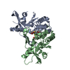
Entry Database : PDB / ID : 5wr7Title Crystal structure of Trk-A complexed with a selective inhibitor CH7057288 High affinity nerve growth factor receptor Keywords / / / / / Function / homology Function Domain/homology Component
/ / / / / / / / / / / / / / / / / / / / / / / / / / / / / / / / / / / / / / / / / / / / / / / / / / / / / / / / / / / / / / / / / / / / / / / / / / / / / / / / / / / / / / / / / / / / / / / / / / / / / / / / / / / / / / / / / / / / / / / / / / / / / / / / / / / / / / Biological species Homo sapiens (human)Method / / / Resolution : 2.76 Å Authors Tanaka, H. / Blaesse, M. / Augustin, M. / Goesser, C. Journal : Mol. Cancer Ther. / Year : 2018Title : Selective TRK Inhibitor CH7057288 against TRK Fusion-Driven Cancer.Authors : Tanaka, H. / Sase, H. / Tsukaguchi, T. / Hasegawa, M. / Tanimura, H. / Yoshida, M. / Sakata, K. / Fujii, T. / Tachibana, Y. / Takanashi, K. / Higashida, A. / Hasegawa, K. / Ono, Y. / Oikawa, N. / Mio, T. History Deposition Nov 30, 2016 Deposition site / Processing site Revision 1.0 Dec 6, 2017 Provider / Type Revision 1.1 Dec 19, 2018 Group / Database references / Category / citation_authorItem _citation.country / _citation.journal_abbrev ... _citation.country / _citation.journal_abbrev / _citation.journal_id_CSD / _citation.journal_id_ISSN / _citation.journal_volume / _citation.page_first / _citation.page_last / _citation.pdbx_database_id_DOI / _citation.pdbx_database_id_PubMed / _citation.title / _citation.year Revision 1.2 Nov 20, 2024 Group / Database references / Structure summaryCategory chem_comp_atom / chem_comp_bond ... chem_comp_atom / chem_comp_bond / database_2 / pdbx_entry_details / pdbx_modification_feature Item / _database_2.pdbx_database_accession
Show all Show less
 Movie
Movie Controller

 Yorodumi
 Open data
 Basic information
 Components
 Keywords
 Function and homology information
 Homo sapiens (human)
 X-RAY DIFFRACTION /
  Authors
 Citation
 Structure visualization
 Downloads & links
 Other downloads
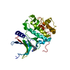


 PDBj
PDBj


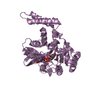
 Assembly
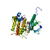

 Spodoptera frugiperda (fall armyworm)
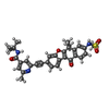
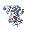
 Mass: 569.671 Da / Num. of mol.: 1 / Source method: obtained synthetically / Formula: C32H31N3O5S
 Mass: 18.015 Da / Num. of mol.: 28 / Source method: isolated from a natural source / Formula: H2O
 Sample preparation
 / Beamline: 08ID-1 / Wavelength: 0.97949 Å
 Processing