 Movie
Movie Controller

[English] 日本語
 Yorodumi
- PDB-5wq8: CryoEM structure of type II secretion system secretin GspD in Vib... -
+ Open data

- Basic information
| Entry | Database: PDB / ID: 5wq8 | ||||||||||||||||||||||||||||||||||||||||||||||||||||||
|---|---|---|---|---|---|---|---|---|---|---|---|---|---|---|---|---|---|---|---|---|---|---|---|---|---|---|---|---|---|---|---|---|---|---|---|---|---|---|---|---|---|---|---|---|---|---|---|---|---|---|---|---|---|---|---|
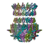
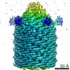
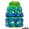
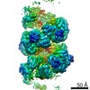
| Title | CryoEM structure of type II secretion system secretin GspD in Vibrio cholerae | ||||||||||||||||||||||||||||||||||||||||||||||||||||||
 Components Components | Type II secretion system protein D | ||||||||||||||||||||||||||||||||||||||||||||||||||||||
 Keywords Keywords | PROTEIN TRANSPORT / Secretin / C15 symmetry / T2SS | ||||||||||||||||||||||||||||||||||||||||||||||||||||||
| Function / homology |  Function and homology information Function and homology informationprotein secretion by the type II secretion system / type II protein secretion system complex / protein secretion / cell outer membrane / identical protein binding Similarity search - Function | ||||||||||||||||||||||||||||||||||||||||||||||||||||||
| Biological species |  Vibrio cholerae O1 biovar El Tor str. N16961 (bacteria) Vibrio cholerae O1 biovar El Tor str. N16961 (bacteria) | ||||||||||||||||||||||||||||||||||||||||||||||||||||||
| Method | ELECTRON MICROSCOPY / single particle reconstruction / cryo EM / Resolution: 3.26 Å | ||||||||||||||||||||||||||||||||||||||||||||||||||||||
 Authors Authors | Yan, Z. / Yin, M. / Li, X. | ||||||||||||||||||||||||||||||||||||||||||||||||||||||
 Citation Citation | Journal: Nat Struct Mol Biol / Year: 2017 Title: Structural insights into the secretin translocation channel in the type II secretion system. Authors: Zhaofeng Yan / Meng Yin / Dandan Xu / Yongqun Zhu / Xueming Li /  Abstract: The secretin GspD of the type II secretion system (T2SS) forms a channel across the outer membrane in Gram-negative bacteria to transport substrates from the periplasm to the extracellular milieu. ...The secretin GspD of the type II secretion system (T2SS) forms a channel across the outer membrane in Gram-negative bacteria to transport substrates from the periplasm to the extracellular milieu. The lack of an atomic-resolution structure of the GspD channel hinders the investigation of substrate translocation mechanism of T2SS. Here we report cryo-EM structures of two GspD channels (∼1 MDa), from Escherichia coli K12 and Vibrio cholerae, at ∼3 Å resolution. The structures reveal a pentadecameric channel architecture, wherein three rings of GspD N domains form the periplasmic channel. The secretin domain constitutes a novel double β-barrel channel, with at least 60 β-strands in each barrel, and is stabilized by S domains. The outer membrane channel is sealed by β-strand-enriched gates. On the basis of the partially open state captured, we proposed a detailed gate-opening mechanism. Our structures provide a structural basis for understanding the secretin superfamily and the mechanism of substrate translocation in T2SS. | ||||||||||||||||||||||||||||||||||||||||||||||||||||||
| History |
|
- Structure visualization
| Movie |
Movie viewer |
|---|---|
| Structure viewer | Molecule: MolmilJmol/JSmol |
- Downloads & links
-Download
| PDBx/mmCIF format | 5wq8.cif.gz | 1.3 MB | Display | PDBx/mmCIF format |
|---|---|---|---|---|
| PDB format | pdb5wq8.ent.gz | 1.1 MB | Display | PDB format |
| PDBx/mmJSON format | 5wq8.json.gz | Tree view | PDBx/mmJSON format | |
| Others |  Other downloads Other downloads |
-Validation report
| Arichive directory | https://data.pdbj.org/pub/pdb/validation_reports/wq/5wq8ftp://data.pdbj.org/pub/pdb/validation_reports/wq/5wq8 | HTTPS FTP |
|---|
-Related structure data
| Related structure data | 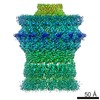 6676MC 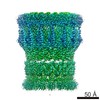 6675C 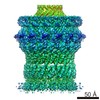 6677C 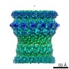 6678C  5wq7C  5wq9C M: map data used to model this data C: citing same article ( |
|---|---|
| Similar structure data |
-Links
 PDBj
PDBj
- Assembly
| Deposited unit | 
|
|---|---|
| 1 |
|
-Components
| #1: Protein | Mass: 70863.078 Da / Num. of mol.: 15 Source method: isolated from a genetically manipulated source Source: (gene. exp.) Vibrio cholerae O1 biovar El Tor str. N16961 (bacteria)Strain: N16961 / Gene: epsD, VC_2733 / Production host: Has protein modification | Y | |
|---|
-Experimental details
-Experiment
| Experiment | Method: ELECTRON MICROSCOPY |
|---|---|
| EM experiment | Aggregation state: PARTICLE / 3D reconstruction method: single particle reconstruction |
- Sample preparation
| Component |
| ||||||||||||||||||
|---|---|---|---|---|---|---|---|---|---|---|---|---|---|---|---|---|---|---|---|
| Molecular weight | Value: 1 MDa / Experimental value: NO | ||||||||||||||||||
| Source (natural) | Organism: Vibrio cholerae O1 (bacteria) | ||||||||||||||||||
| Source (recombinant) | Organism: | ||||||||||||||||||
| Buffer solution | pH: 8 | ||||||||||||||||||
| Specimen | Embedding applied: NO / Shadowing applied: NO / Staining applied: NO / Vitrification applied: YES | ||||||||||||||||||
| Vitrification | Cryogen name: ETHANE |
- Electron microscopy imaging
| Experimental equipment |  Model: Titan Krios / Image courtesy: FEI Company |
|---|---|
| Microscopy | Model: FEI TITAN KRIOS |
| Electron gun | Electron source:  FIELD EMISSION GUN / Accelerating voltage: 300 kV / Illumination mode: OTHER FIELD EMISSION GUN / Accelerating voltage: 300 kV / Illumination mode: OTHER |
| Electron lens | Mode: BRIGHT FIELD |
| Image recording | Electron dose: 1.6 e/Å2 / Film or detector model: GATAN K2 SUMMIT (4k x 4k) |
- Processing
| Software | Name: PHENIX / Version: 1.10_2152: / Classification: refinement | ||||||||||||||||||||||||
|---|---|---|---|---|---|---|---|---|---|---|---|---|---|---|---|---|---|---|---|---|---|---|---|---|---|
| EM software |
| ||||||||||||||||||||||||
| CTF correction | Type: PHASE FLIPPING AND AMPLITUDE CORRECTION | ||||||||||||||||||||||||
| Symmetry | Point symmetry: C15 (15 fold cyclic) | ||||||||||||||||||||||||
| 3D reconstruction | Resolution: 3.26 Å / Resolution method: FSC 0.143 CUT-OFF / Num. of particles: 41579 / Symmetry type: POINT | ||||||||||||||||||||||||
| Refine LS restraints |
|
