 Movie
Movie Controller

[English] 日本語
 Yorodumi
- PDB-5wq2: Crystal structure of 3C protease from a mild Human enterovirus 71... -
+ Open data

- Basic information
| Entry | Database: PDB / ID: 5wq2 | ||||||
|---|---|---|---|---|---|---|---|
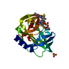
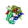
| Title | Crystal structure of 3C protease from a mild Human enterovirus 71 in complex with rupintrivir | ||||||
 Components Components | 3C protein | ||||||
 Keywords Keywords | HYDROLASE / 3C / mutation / virus | ||||||
| Function / homology |  Function and homology information Function and homology informationT=pseudo3 icosahedral viral capsid / host cell cytoplasm / cysteine-type endopeptidase activity / symbiont entry into host cell / virion attachment to host cell / proteolysis / RNA binding Similarity search - Function | ||||||
| Biological species |   Enterovirus A71 Enterovirus A71 | ||||||
| Method |  X-RAY DIFFRACTION / SYNCHROTRON / MOLECULAR REPLACEMENT / Resolution: 1.39 Å X-RAY DIFFRACTION / SYNCHROTRON / MOLECULAR REPLACEMENT / Resolution: 1.39 Å | ||||||
 Authors Authors | Li, B. / Yuan, Z. | ||||||
 Citation Citation | Journal: To Be Published Title: Crystal structure of 3C protease from a mild Human enterovirus 71 in complex with rupintrivir Authors: Li, B. / Qin, L. | ||||||
| History |
|
- Structure visualization
| Structure viewer | Molecule: MolmilJmol/JSmol |
|---|
- Downloads & links
-Download
| PDBx/mmCIF format | 5wq2.cif.gz | 59.4 KB | Display | PDBx/mmCIF format |
|---|---|---|---|---|
| PDB format | pdb5wq2.ent.gz | 41.5 KB | Display | PDB format |
| PDBx/mmJSON format | 5wq2.json.gz | Tree view | PDBx/mmJSON format | |
| Others |  Other downloads Other downloads |
-Validation report
| Arichive directory | https://data.pdbj.org/pub/pdb/validation_reports/wq/5wq2ftp://data.pdbj.org/pub/pdb/validation_reports/wq/5wq2 | HTTPS FTP |
|---|
-Related structure data


-Links
 PDBj
PDBj

- Assembly
| Deposited unit | 
| ||||||||
|---|---|---|---|---|---|---|---|---|---|
| 1 |
| ||||||||
| Unit cell |
|
-Components
| #1: Protein | Mass: 20092.123 Da / Num. of mol.: 1 Source method: isolated from a genetically manipulated source Source: (gene. exp.) Enterovirus A71 / Production host:  | ||
|---|---|---|---|
| #2: Chemical | ChemComp-AG7 /   Mass: 600.678 Da / Num. of mol.: 1 / Source method: obtained synthetically / Formula: C31H41FN4O7 / Comment: antivirus, protease inhibitor*YM Mass: 600.678 Da / Num. of mol.: 1 / Source method: obtained synthetically / Formula: C31H41FN4O7 / Comment: antivirus, protease inhibitor*YM | ||
| #3: Chemical |   Mass: 96.063 Da / Num. of mol.: 2 / Source method: obtained synthetically / Formula: SO4 Mass: 96.063 Da / Num. of mol.: 2 / Source method: obtained synthetically / Formula: SO4#4: Water | ChemComp-HOH / |  Mass: 18.015 Da / Num. of mol.: 236 / Source method: isolated from a natural source / Formula: H2O Mass: 18.015 Da / Num. of mol.: 236 / Source method: isolated from a natural source / Formula: H2O |
-Experimental details
-Experiment
| Experiment | Method: X-RAY DIFFRACTION / Number of used crystals: 1 |
|---|
- Sample preparation
| Crystal | Density Matthews: 2.06 Å3/Da / Density % sol: 40.24 % |
|---|---|
| Crystal grow | Temperature: 289 K / Method: vapor diffusion, hanging drop / Details: 28% PEG 8000, Acetate PH4.5, 0.2M Li2SO4 |
-Data collection
| Diffraction | Mean temperature: 100 K |
|---|---|
| Diffraction source | Source: SYNCHROTRON / Site: SSRF  / Beamline: BL17U / Wavelength: 0.9798 Å / Beamline: BL17U / Wavelength: 0.9798 Å |
| Detector | Type: ADSC QUANTUM 315r / Detector: CCD / Date: Jan 15, 2015 |
| Radiation | Protocol: SINGLE WAVELENGTH / Monochromatic (M) / Laue (L): M / Scattering type: x-ray |
| Radiation wavelength | Wavelength: 0.9798 Å / Relative weight: 1 |
| Reflection | Resolution: 1.39→50 Å / Num. obs: 32549 / % possible obs: 99 % / Redundancy: 3.7 % / Net I/σ(I): 22.38 |
- Processing
| Software |
| |||||||||||||||||||||||||||||||||||||||||||||||||||||||||||||||||||||||||||||||||||||||||||||||||||||||||
|---|---|---|---|---|---|---|---|---|---|---|---|---|---|---|---|---|---|---|---|---|---|---|---|---|---|---|---|---|---|---|---|---|---|---|---|---|---|---|---|---|---|---|---|---|---|---|---|---|---|---|---|---|---|---|---|---|---|---|---|---|---|---|---|---|---|---|---|---|---|---|---|---|---|---|---|---|---|---|---|---|---|---|---|---|---|---|---|---|---|---|---|---|---|---|---|---|---|---|---|---|---|---|---|---|---|---|
| Refinement | Method to determine structure: MOLECULAR REPLACEMENT / Resolution: 1.39→23.224 Å / SU ML: 0.14 / Cross valid method: FREE R-VALUE / σ(F): 1.35 / Phase error: 21.14
| |||||||||||||||||||||||||||||||||||||||||||||||||||||||||||||||||||||||||||||||||||||||||||||||||||||||||
| Solvent computation | Shrinkage radii: 0.9 Å / VDW probe radii: 1.11 Å | |||||||||||||||||||||||||||||||||||||||||||||||||||||||||||||||||||||||||||||||||||||||||||||||||||||||||
| Refinement step | Cycle: LAST / Resolution: 1.39→23.224 Å
| |||||||||||||||||||||||||||||||||||||||||||||||||||||||||||||||||||||||||||||||||||||||||||||||||||||||||
| Refine LS restraints |
| |||||||||||||||||||||||||||||||||||||||||||||||||||||||||||||||||||||||||||||||||||||||||||||||||||||||||
| LS refinement shell |
|
