 Movie
Movie Controller


+ Open data

- Basic information
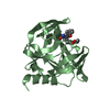
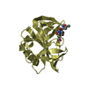
| Entry | Database: PDB / ID: 5gsw | ||||||
|---|---|---|---|---|---|---|---|
| Title | Crystal structure of EV71 3C in complex with N69S 1.8k | ||||||
 Components Components | 3C protein | ||||||
 Keywords Keywords | HYDROLASE / Protease | ||||||
| Function / homology |  Function and homology information Function and homology informationT=pseudo3 icosahedral viral capsid / host cell cytoplasm / cysteine-type endopeptidase activity / symbiont entry into host cell / virion attachment to host cell / proteolysis / RNA binding Similarity search - Function | ||||||
| Biological species |   Enterovirus A71 Enterovirus A71 | ||||||
| Method |  X-RAY DIFFRACTION / MOLECULAR REPLACEMENT / Resolution: 3.19 Å X-RAY DIFFRACTION / MOLECULAR REPLACEMENT / Resolution: 3.19 Å | ||||||
 Authors Authors | Wang, Y. | ||||||
| Funding support |  China, 1items China, 1items
| ||||||
 Citation Citation | Journal: Antimicrob. Agents Chemother. / Year: 2017 Title: Structure of the Enterovirus 71 3C Protease in Complex with NK-1.8k and Indications for the Development of Antienterovirus Protease Inhibitor Authors: Wang, Y. / Cao, L. / Zhai, Y. / Yin, Z. / Sun, Y. / Shang, L. | ||||||
| History |
|
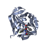
- Structure visualization
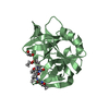
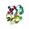
| Structure viewer | Molecule: MolmilJmol/JSmol |
|---|
- Downloads & links
-Download
| PDBx/mmCIF format | 5gsw.cif.gz | 357.8 KB | Display | PDBx/mmCIF format |
|---|---|---|---|---|
| PDB format | pdb5gsw.ent.gz | 296.1 KB | Display | PDB format |
| PDBx/mmJSON format | 5gsw.json.gz | Tree view | PDBx/mmJSON format | |
| Others |  Other downloads Other downloads |
-Validation report
| Arichive directory | https://data.pdbj.org/pub/pdb/validation_reports/gs/5gswftp://data.pdbj.org/pub/pdb/validation_reports/gs/5gsw | HTTPS FTP |
|---|
-Related structure data
| Related structure data |  5gsoC 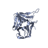 3osyS C: citing same article ( S: Starting model for refinement |
|---|---|
| Similar structure data |


-Links
 PDBj
PDBj

- Assembly
| Deposited unit | 
| ||||||||
|---|---|---|---|---|---|---|---|---|---|
| 1 | 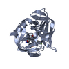
| ||||||||
| 2 | 
| ||||||||
| 3 | 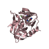
| ||||||||
| 4 | 
| ||||||||
| 5 | 
| ||||||||
| Unit cell |
|
-Components
| #1: Protein | Mass: 20162.107 Da / Num. of mol.: 5 / Mutation: N69S Source method: isolated from a genetically manipulated source Source: (gene. exp.) Enterovirus A71Production host: References: UniProt: E7E815 #2: Chemical | ChemComp-5GI / ~{   Mass: 444.456 Da / Num. of mol.: 5 Mass: 444.456 Da / Num. of mol.: 5Source method: isolated from a genetically manipulated source Formula: C22H25FN4O5 #3: Water | ChemComp-HOH / |  Mass: 18.015 Da / Num. of mol.: 31 / Source method: isolated from a natural source / Formula: H2O Mass: 18.015 Da / Num. of mol.: 31 / Source method: isolated from a natural source / Formula: H2O |
|---|
-Experimental details
-Experiment
| Experiment | Method: X-RAY DIFFRACTION / Number of used crystals: 1 |
|---|
- Sample preparation
| Crystal | Density Matthews: 2.71 Å3/Da / Density % sol: 54.57 % |
|---|---|
| Crystal grow | Temperature: 289 K / Method: vapor diffusion, hanging drop / pH: 7.7 Details: 0.1 M Tris-HCl, pH 7.7, 200 mM Sodium Citrate, 16% PEG 3350. |
-Data collection
| Diffraction | Mean temperature: 100 K |
|---|---|
| Diffraction source | Source: ROTATING ANODE / Type: Cu FINE FOCUS / Wavelength: 1.5418 Å |
| Detector | Type: RIGAKU SATURN 944 / Detector: CCD / Date: May 8, 2015 |
| Radiation | Protocol: SINGLE WAVELENGTH / Monochromatic (M) / Laue (L): M / Scattering type: x-ray |
| Radiation wavelength | Wavelength: 1.5418 Å / Relative weight: 1 |
| Reflection | Resolution: 3.2→50 Å / Num. obs: 17983 / % possible obs: 96.8 % / Redundancy: 1.7 % / Rmerge(I) obs: 0.157 / Net I/σ(I): 7.36 |
| Reflection shell | Resolution: 3.2→8.67 Å / Rmerge(I) obs: 0.581 |
- Processing
| Software |
| ||||||||||||||||||||||||||||||||||||||||||||||||||||||||||||||||||||||||||||||||||||||||||||||||||||||||||||||||||||||||||||||||||||||||||||||||||||||
|---|---|---|---|---|---|---|---|---|---|---|---|---|---|---|---|---|---|---|---|---|---|---|---|---|---|---|---|---|---|---|---|---|---|---|---|---|---|---|---|---|---|---|---|---|---|---|---|---|---|---|---|---|---|---|---|---|---|---|---|---|---|---|---|---|---|---|---|---|---|---|---|---|---|---|---|---|---|---|---|---|---|---|---|---|---|---|---|---|---|---|---|---|---|---|---|---|---|---|---|---|---|---|---|---|---|---|---|---|---|---|---|---|---|---|---|---|---|---|---|---|---|---|---|---|---|---|---|---|---|---|---|---|---|---|---|---|---|---|---|---|---|---|---|---|---|---|---|---|---|---|---|
| Refinement | Method to determine structure: MOLECULAR REPLACEMENT Starting model: 3OSY Resolution: 3.19→48.62 Å / SU ML: 0.48 / Cross valid method: NONE / σ(F): 1.34 / Phase error: 26.93
| ||||||||||||||||||||||||||||||||||||||||||||||||||||||||||||||||||||||||||||||||||||||||||||||||||||||||||||||||||||||||||||||||||||||||||||||||||||||
| Solvent computation | Shrinkage radii: 0.9 Å / VDW probe radii: 1.11 Å | ||||||||||||||||||||||||||||||||||||||||||||||||||||||||||||||||||||||||||||||||||||||||||||||||||||||||||||||||||||||||||||||||||||||||||||||||||||||
| Refinement step | Cycle: LAST / Resolution: 3.19→48.62 Å
| ||||||||||||||||||||||||||||||||||||||||||||||||||||||||||||||||||||||||||||||||||||||||||||||||||||||||||||||||||||||||||||||||||||||||||||||||||||||
| Refine LS restraints |
| ||||||||||||||||||||||||||||||||||||||||||||||||||||||||||||||||||||||||||||||||||||||||||||||||||||||||||||||||||||||||||||||||||||||||||||||||||||||
| LS refinement shell |
| ||||||||||||||||||||||||||||||||||||||||||||||||||||||||||||||||||||||||||||||||||||||||||||||||||||||||||||||||||||||||||||||||||||||||||||||||||||||
| Refinement TLS params. | Method: refined / Refine-ID: X-RAY DIFFRACTION
| ||||||||||||||||||||||||||||||||||||||||||||||||||||||||||||||||||||||||||||||||||||||||||||||||||||||||||||||||||||||||||||||||||||||||||||||||||||||
| Refinement TLS group |
|
