 Movie
Movie Controller


+ Open data

- Basic information
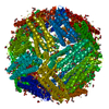
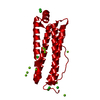
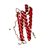
| Entry | Database: PDB / ID: 5wpn | ||||||
|---|---|---|---|---|---|---|---|
| Title | Zn-bound Structure of Chaetopterus variopedatus Ferritin | ||||||
 Components Components | Ferritin | ||||||
 Keywords Keywords | OXIDOREDUCTASE / Ferritin | ||||||
| Function / homology |  Function and homology information Function and homology information: / ferroxidase / ferroxidase activity / ferric iron binding / iron ion transport / ferrous iron binding / cytoplasm Similarity search - Function | ||||||
| Biological species |  Chaetopterus variopedatus (invertebrata) Chaetopterus variopedatus (invertebrata) | ||||||
| Method |  X-RAY DIFFRACTION / SYNCHROTRON / MOLECULAR REPLACEMENT / Resolution: 1.57 Å X-RAY DIFFRACTION / SYNCHROTRON / MOLECULAR REPLACEMENT / Resolution: 1.57 Å | ||||||
 Authors Authors | De Meulenaere, E. / Bailey, J.B. / Tezcan, F.A. / Deheyn, D. | ||||||
 Citation Citation | Journal: Biochem. J. / Year: 2017 Title: First biochemical and crystallographic characterization of a fast-performing ferritin from a marine invertebrate. Authors: De Meulenaere, E. / Bailey, J.B. / Tezcan, F.A. / Deheyn, D.D. | ||||||
| History |
|
- Structure visualization
| Structure viewer | Molecule: MolmilJmol/JSmol |
|---|
- Downloads & links
-Download
| PDBx/mmCIF format | 5wpn.cif.gz | 91.5 KB | Display | PDBx/mmCIF format |
|---|---|---|---|---|
| PDB format | pdb5wpn.ent.gz | 71.2 KB | Display | PDB format |
| PDBx/mmJSON format | 5wpn.json.gz | Tree view | PDBx/mmJSON format | |
| Others |  Other downloads Other downloads |
-Validation report
| Arichive directory | https://data.pdbj.org/pub/pdb/validation_reports/wp/5wpnftp://data.pdbj.org/pub/pdb/validation_reports/wp/5wpn | HTTPS FTP |
|---|
-Related structure data
| Similar structure data |
|---|


-Links
 PDBj
PDBj


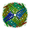
- Assembly
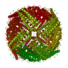
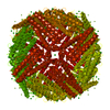
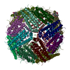
| Deposited unit | 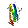
| ||||||||||||||||||||||||||||||
|---|---|---|---|---|---|---|---|---|---|---|---|---|---|---|---|---|---|---|---|---|---|---|---|---|---|---|---|---|---|---|---|
| 1 | x 24
| ||||||||||||||||||||||||||||||
| Unit cell |
| ||||||||||||||||||||||||||||||
| Components on special symmetry positions |
| ||||||||||||||||||||||||||||||
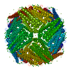
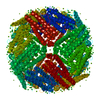
| Details | 24-meric cage-like protein by gel filtration |
-Components
-Protein , 1 types, 1 molecules A
| #1: Protein | Mass: 19824.100 Da / Num. of mol.: 1 / Mutation: N82D Source method: isolated from a genetically manipulated source Source: (gene. exp.) Chaetopterus variopedatus (invertebrata)Production host:  |
|---|
-Non-polymers , 6 types, 305 molecules 


| #2: Chemical | ChemComp-ZN /  Mass: 65.409 Da / Num. of mol.: 12 / Source method: obtained synthetically / Formula: Zn Mass: 65.409 Da / Num. of mol.: 12 / Source method: obtained synthetically / Formula: Zn#3: Chemical | ChemComp-CA / |  Mass: 40.078 Da / Num. of mol.: 1 / Source method: obtained synthetically / Formula: Ca Mass: 40.078 Da / Num. of mol.: 1 / Source method: obtained synthetically / Formula: Ca#4: Chemical | ChemComp-CL / |  Mass: 35.453 Da / Num. of mol.: 1 / Source method: obtained synthetically / Formula: Cl Mass: 35.453 Da / Num. of mol.: 1 / Source method: obtained synthetically / Formula: Cl#5: Chemical |  Mass: 106.120 Da / Num. of mol.: 2 / Source method: obtained synthetically / Formula: C4H10O3 Mass: 106.120 Da / Num. of mol.: 2 / Source method: obtained synthetically / Formula: C4H10O3#6: Chemical | ChemComp-EDO / |  Mass: 62.068 Da / Num. of mol.: 1 / Source method: obtained synthetically / Formula: C2H6O2 Mass: 62.068 Da / Num. of mol.: 1 / Source method: obtained synthetically / Formula: C2H6O2#7: Water | ChemComp-HOH / | Mass: 18.015 Da / Num. of mol.: 288 / Source method: isolated from a natural source / Formula: H2O |
|---|
-Experimental details
-Experiment
| Experiment | Method: X-RAY DIFFRACTION / Number of used crystals: 1 |
|---|
- Sample preparation
| Crystal | Density Matthews: 3.19 Å3/Da / Density % sol: 61.4 % |
|---|---|
| Crystal grow | Temperature: 297 K / Method: vapor diffusion, sitting drop Details: Reservoir: 500 uL total volume: 20 mM Tris (pH 8.5), 40 mM CaCl2, 6% PEG 400 Sitting Drop: 2 uL reservoir, 2 uL of 25 uM Chaetopterus variopedatus ferritin Soaking Solution (30 min): 10 mM ...Details: Reservoir: 500 uL total volume: 20 mM Tris (pH 8.5), 40 mM CaCl2, 6% PEG 400 Sitting Drop: 2 uL reservoir, 2 uL of 25 uM Chaetopterus variopedatus ferritin Soaking Solution (30 min): 10 mM Zn, 20 mM Tris (pH 8.5), 20 mM CaCl2, and 3% (v/v) PEG 400 |
-Data collection
| Diffraction | Mean temperature: 100 K |
|---|---|
| Diffraction source | Source: SYNCHROTRON / Site: SSRL  / Beamline: BL12-2 / Wavelength: 0.97946 / Beamline: BL12-2 / Wavelength: 0.97946 |
| Detector | Type: DECTRIS PILATUS 6M / Detector: PIXEL / Date: Jan 14, 2016 |
| Radiation | Protocol: SINGLE WAVELENGTH / Monochromatic (M) / Laue (L): M / Scattering type: x-ray / Wavelength: 0.97946 |
| Radiation wavelength | Wavelength: 0.97946 Å / Relative weight: 1 |
| Reflection | Resolution: 1.57→90.94 Å / Num. obs: 36446 / % possible obs: 99.9 % / Redundancy: 25.1 % / CC1/2: 0.985 / Rmerge(I) obs: 0.095 / Net I/σ(I): 24.9 |
| Reflection shell | Resolution: 1.57→1.6 Å / Redundancy: 5.6 % / Rmerge(I) obs: 0.803 / Mean I/σ(I) obs: 2.2 / Num. unique obs: 296 / CC1/2: 0.568 / % possible all: 98.6 |
- Processing
| Software |
| ||||||||||||||||||||
|---|---|---|---|---|---|---|---|---|---|---|---|---|---|---|---|---|---|---|---|---|---|
| Refinement | Method to determine structure: MOLECULAR REPLACEMENT Starting model: homology model Resolution: 1.57→45.467 Å / Cross valid method: THROUGHOUT
| ||||||||||||||||||||
| Solvent computation | Shrinkage radii: 0.9 Å / VDW probe radii: 1.11 Å | ||||||||||||||||||||
| Refinement step | Cycle: LAST / Resolution: 1.57→45.467 Å
| ||||||||||||||||||||
| LS refinement shell | Resolution: 1.57→1.61 Å /
|
