 Movie
Movie Controller

[English] 日本語
 Yorodumi
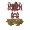
- PDB-5wie: Crystal structure of a Kv1.2-2.1 chimera K+ channel V406W mutant ... -
+ Open data

- Basic information
| Entry | Database: PDB / ID: 5wie | |||||||||
|---|---|---|---|---|---|---|---|---|---|---|
| Title | Crystal structure of a Kv1.2-2.1 chimera K+ channel V406W mutant in an inactivated state | |||||||||
 Components Components |
| |||||||||
 Keywords Keywords | MEMBRANE PROTEIN / METAL TRANSPORT / Ion Channel / Inactivation / Voltage-gated | |||||||||
| Function / homology |  Function and homology information Function and homology informationVoltage gated Potassium channels / optic nerve structural organization / pinceau fiber / methylglyoxal reductase (NADPH) (acetol producing) activity / Voltage gated Potassium channels / potassium channel complex / voltage-gated monoatomic ion channel activity involved in regulation of postsynaptic membrane potential / regulation of circadian sleep/wake cycle, non-REM sleep / paranodal junction / potassium ion export across plasma membrane ...Voltage gated Potassium channels / optic nerve structural organization / pinceau fiber / methylglyoxal reductase (NADPH) (acetol producing) activity / Voltage gated Potassium channels / potassium channel complex / voltage-gated monoatomic ion channel activity involved in regulation of postsynaptic membrane potential / regulation of circadian sleep/wake cycle, non-REM sleep / paranodal junction / potassium ion export across plasma membrane / regulation of protein localization to cell surface / corpus callosum development / : / voltage-gated monoatomic ion channel activity involved in regulation of presynaptic membrane potential / delayed rectifier potassium channel activity / axon initial segment / optic nerve development / Oxidoreductases; Acting on the CH-OH group of donors; With NAD+ or NADP+ as acceptor / juxtaparanode region of axon / outward rectifier potassium channel activity / myoblast differentiation / regulation of potassium ion transmembrane transport / Neutrophil degranulation / neuronal cell body membrane / neuromuscular process / regulation of dopamine secretion / lamellipodium membrane / action potential / voltage-gated potassium channel activity / kinesin binding / potassium channel regulator activity / hematopoietic progenitor cell differentiation / neuronal action potential / voltage-gated potassium channel complex / axon terminus / potassium ion transmembrane transport / sensory perception of pain / calyx of Held / postsynaptic density membrane / protein homooligomerization / cerebral cortex development / cytoplasmic side of plasma membrane / lamellipodium / presynaptic membrane / perikaryon / transmembrane transporter binding / postsynaptic membrane / cytoskeleton / endosome / neuron projection / postsynaptic density / axon / dendrite / endoplasmic reticulum membrane / protein-containing complex binding / glutamatergic synapse / membrane / plasma membrane / cytosol Similarity search - Function | |||||||||
| Biological species |  | |||||||||
| Method |  X-RAY DIFFRACTION / SYNCHROTRON / MOLECULAR REPLACEMENT / Resolution: 3.3 Å X-RAY DIFFRACTION / SYNCHROTRON / MOLECULAR REPLACEMENT / Resolution: 3.3 Å | |||||||||
 Authors Authors | Pau, V. / Zhou, Y. / Ramu, Y. / Xu, Y. / Lu, Z. | |||||||||
| Funding support |  United States, 2items United States, 2items
| |||||||||
 Citation Citation | Journal: Nat. Struct. Mol. Biol. / Year: 2017 Title: Crystal structure of an inactivated mutant mammalian voltage-gated K(+) channel. Authors: Pau, V. / Zhou, Y. / Ramu, Y. / Xu, Y. / Lu, Z. | |||||||||
| History |
|
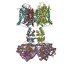
- Structure visualization
| Structure viewer | Molecule: MolmilJmol/JSmol |
|---|
- Downloads & links
-Download
| PDBx/mmCIF format | 5wie.cif.gz | 296.5 KB | Display | PDBx/mmCIF format |
|---|---|---|---|---|
| PDB format | pdb5wie.ent.gz | 228.2 KB | Display | PDB format |
| PDBx/mmJSON format | 5wie.json.gz | Tree view | PDBx/mmJSON format | |
| Others |  Other downloads Other downloads |
-Validation report
| Summary document | 5wie_validation.pdf.gz | 1.9 MB | Display | wwPDB validaton report |
|---|---|---|---|---|
| Full document | 5wie_full_validation.pdf.gz | 1.9 MB | Display | |
| Data in XML | 5wie_validation.xml.gz | 50.6 KB | Display | |
| Data in CIF | 5wie_validation.cif.gz | 69.3 KB | Display | |
| Arichive directory | https://data.pdbj.org/pub/pdb/validation_reports/wi/5wieftp://data.pdbj.org/pub/pdb/validation_reports/wi/5wie | HTTPS FTP |
-Related structure data
| Related structure data | 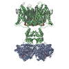 2r9rS S: Starting model for refinement |
|---|---|
| Similar structure data |

-Links
 PDBj
PDBj


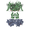
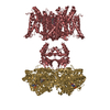
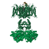
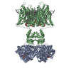
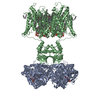
- Assembly
| Deposited unit | 
| |||||||||||||||||||||||||||
|---|---|---|---|---|---|---|---|---|---|---|---|---|---|---|---|---|---|---|---|---|---|---|---|---|---|---|---|---|
| 1 | 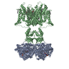
| |||||||||||||||||||||||||||
| 2 | 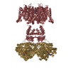
| |||||||||||||||||||||||||||
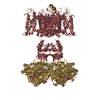
| Unit cell |
| |||||||||||||||||||||||||||
| Components on special symmetry positions |
|
-Components
| #1: Protein | Mass: 37353.086 Da / Num. of mol.: 2 Source method: isolated from a genetically manipulated source Source: (gene. exp.)  Pichia (fungus) Pichia (fungus)References: UniProt: P62483, Oxidoreductases; Acting on the CH-OH group of donors; With NAD+ or NADP+ as acceptor #2: Protein | Mass: 60455.312 Da / Num. of mol.: 2 / Mutation: V406W Source method: isolated from a genetically manipulated source Source: (gene. exp.) Gene: Kcna2 / Plasmid: pPICZ-C / Production host: Pichia (fungus) / References: UniProt: P63142, UniProt: P63141#3: Chemical |   Mass: 743.405 Da / Num. of mol.: 2 / Source method: obtained synthetically / Formula: C21H28N7O17P3 Mass: 743.405 Da / Num. of mol.: 2 / Source method: obtained synthetically / Formula: C21H28N7O17P3#4: Chemical | ChemComp-PGW / ( 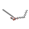  Mass: 749.007 Da / Num. of mol.: 6 / Source method: obtained synthetically / Formula: C40H77O10P / Comment: phospholipid*YM Mass: 749.007 Da / Num. of mol.: 6 / Source method: obtained synthetically / Formula: C40H77O10P / Comment: phospholipid*YM#5: Chemical | ChemComp-K /   Mass: 39.098 Da / Num. of mol.: 8 / Source method: obtained synthetically / Formula: K Mass: 39.098 Da / Num. of mol.: 8 / Source method: obtained synthetically / Formula: K |
|---|
-Experimental details
-Experiment
| Experiment | Method: X-RAY DIFFRACTION / Number of used crystals: 1 |
|---|
- Sample preparation
| Crystal | Density Matthews: 3.72 Å3/Da / Density % sol: 73.59 % |
|---|---|
| Crystal grow | Temperature: 293 K / Method: vapor diffusion / pH: 8 / Details: 50 mM Tris-Cl pH 8.3, 29-31% PEG400 |
-Data collection
| Diffraction | Mean temperature: 100 K |
|---|---|
| Diffraction source | Source: SYNCHROTRON / Site: ALS / Beamline: 8.2.1 / Wavelength: 1 Å |
| Detector | Type: ADSC QUANTUM 315r / Detector: CCD / Date: Apr 17, 2014 |
| Radiation | Protocol: SINGLE WAVELENGTH / Monochromatic (M) / Laue (L): M / Scattering type: x-ray |
| Radiation wavelength | Wavelength: 1 Å / Relative weight: 1 |
| Reflection | Resolution: 3.3→50 Å / Num. obs: 45103 / % possible obs: 99.5 % / Redundancy: 11.7 % / CC1/2: 0.985 / Rmerge(I) obs: 0.33 / Rpim(I) all: 0.11 / Net I/σ(I): 7.9 |
| Reflection shell | Resolution: 3.3→3.42 Å / Redundancy: 9 % / Mean I/σ(I) obs: 1.7 / CC1/2: 0.604 / Rpim(I) all: 0.413 / % possible all: 97.2 |
- Processing
| Software |
| ||||||||||||||||
|---|---|---|---|---|---|---|---|---|---|---|---|---|---|---|---|---|---|
| Refinement | Method to determine structure: MOLECULAR REPLACEMENT Starting model: 2R9R Resolution: 3.3→40 Å / Cross valid method: FREE R-VALUE
| ||||||||||||||||
| Refinement step | Cycle: LAST / Resolution: 3.3→40 Å
|
