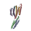
 Movie
Movie Controller


+ Open data

- Basic information
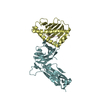
| Entry | Database: PDB / ID: 5we1 | ||||||
|---|---|---|---|---|---|---|---|
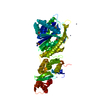
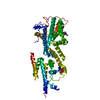
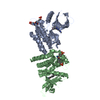
| Title | Structural Basis for Shelterin Bridge Assembly | ||||||
 Components Components |
| ||||||
 Keywords Keywords | GENE REGULATION / Telomere / Shelterin / Cooperativity | ||||||
| Function / homology |  Function and homology information Function and homology informationtelomere-telomerase complex assembly / telomere cap complex / chromosome, telomeric repeat region / telomere maintenance via telomere lengthening / shelterin complex / telomere capping / telomerase holoenzyme complex / telomeric repeat DNA binding / telomere maintenance via telomerase / telomere organization ...telomere-telomerase complex assembly / telomere cap complex / chromosome, telomeric repeat region / telomere maintenance via telomere lengthening / shelterin complex / telomere capping / telomerase holoenzyme complex / telomeric repeat DNA binding / telomere maintenance via telomerase / telomere organization / telomere maintenance / DNA binding / nucleus / cytosol Similarity search - Function | ||||||
| Biological species |  | ||||||
| Method |  X-RAY DIFFRACTION / SYNCHROTRON / MOLECULAR REPLACEMENT / Resolution: 3.202 Å X-RAY DIFFRACTION / SYNCHROTRON / MOLECULAR REPLACEMENT / Resolution: 3.202 Å | ||||||
 Authors Authors | Kim, J.-K. / Liu, J. / Hu, X. / Yu, C. / Roskamp, K. / Sankaran, B. / Huang, L. / Komives, E.-A. / Qiao, F. | ||||||
 Citation Citation | Journal: Mol. Cell / Year: 2017 Title: Structural Basis for Shelterin Bridge Assembly. Authors: Kim, J.K. / Liu, J. / Hu, X. / Yu, C. / Roskamp, K. / Sankaran, B. / Huang, L. / Komives, E.A. / Qiao, F. | ||||||
| History |
|
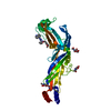
- Structure visualization
| Structure viewer | Molecule: MolmilJmol/JSmol |
|---|
- Downloads & links
-Download
| PDBx/mmCIF format | 5we1.cif.gz | 100.8 KB | Display | PDBx/mmCIF format |
|---|---|---|---|---|
| PDB format | pdb5we1.ent.gz | 76.5 KB | Display | PDB format |
| PDBx/mmJSON format | 5we1.json.gz | Tree view | PDBx/mmJSON format | |
| Others |  Other downloads Other downloads |
-Validation report
| Arichive directory | https://data.pdbj.org/pub/pdb/validation_reports/we/5we1ftp://data.pdbj.org/pub/pdb/validation_reports/we/5we1 | HTTPS FTP |
|---|
-Related structure data
| Related structure data |  5we0C  5we2SC S: Starting model for refinement C: citing same article ( |
|---|---|
| Similar structure data |


-Links
 PDBj
PDBj
- Assembly
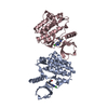
| Deposited unit | 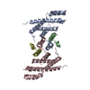
| ||||||||
|---|---|---|---|---|---|---|---|---|---|
| 1 |
| ||||||||
| Unit cell |
|
-Components
| #1: Protein | Mass: 25314.229 Da / Num. of mol.: 2 Source method: isolated from a genetically manipulated source Source: (gene. exp.) Strain: 972 / ATCC 24843 / Gene: poz1, SPAC19G12.13c / Production host:  #2: Protein/peptide | Mass: 3964.574 Da / Num. of mol.: 2 / Fragment: UNP residues 476-508 Source method: isolated from a genetically manipulated source Source: (gene. exp.) Strain: 972 / ATCC 24843 / Gene: tpz1, mug169, SPAC6F6.16c, SPAC6F6.18c / Production host: #3: Chemical |   Mass: 65.409 Da / Num. of mol.: 2 / Source method: obtained synthetically / Formula: Zn Mass: 65.409 Da / Num. of mol.: 2 / Source method: obtained synthetically / Formula: ZnHas protein modification | Y | |
|---|
-Experimental details
-Experiment
| Experiment | Method: X-RAY DIFFRACTION / Number of used crystals: 1 |
|---|
- Sample preparation
| Crystal | Density Matthews: 2.67 Å3/Da / Density % sol: 53.93 % |
|---|---|
| Crystal grow | Temperature: 291 K / Method: vapor diffusion, hanging drop Details: 0.1 M Tris-HCl pH 8.0, 4% Reagent Alcohol, 0.3 M MgCl2 |
-Data collection
| Diffraction | Mean temperature: 100 K |
|---|---|
| Diffraction source | Source: SYNCHROTRON / Site: ALS  / Beamline: 5.0.1 / Wavelength: 0.977408 Å / Beamline: 5.0.1 / Wavelength: 0.977408 Å |
| Detector | Type: ADSC QUANTUM 315r / Detector: CCD / Date: Apr 28, 2016 |
| Radiation | Protocol: SINGLE WAVELENGTH / Monochromatic (M) / Laue (L): M / Scattering type: x-ray |
| Radiation wavelength | Wavelength: 0.977408 Å / Relative weight: 1 |
| Reflection | Resolution: 3.2→57.25 Å / Num. obs: 9911 / % possible obs: 99.7 % / Redundancy: 5.3 % / Rmerge(I) obs: 0.104 / Net I/σ(I): 11.5 |
| Reflection shell | Resolution: 3.2→3.37 Å / Redundancy: 5.4 % / Rmerge(I) obs: 0.765 / Mean I/σ(I) obs: 2.6 / % possible all: 99.5 |
- Processing
| Software |
| |||||||||||||||||||||||||||||||||||
|---|---|---|---|---|---|---|---|---|---|---|---|---|---|---|---|---|---|---|---|---|---|---|---|---|---|---|---|---|---|---|---|---|---|---|---|---|
| Refinement | Method to determine structure: MOLECULAR REPLACEMENT Starting model: 5WE2 Resolution: 3.202→51.917 Å / SU ML: 0.56 / Cross valid method: FREE R-VALUE / σ(F): 2 / Phase error: 40.96
| |||||||||||||||||||||||||||||||||||
| Solvent computation | Shrinkage radii: 0.9 Å / VDW probe radii: 1.11 Å | |||||||||||||||||||||||||||||||||||
| Refinement step | Cycle: LAST / Resolution: 3.202→51.917 Å
| |||||||||||||||||||||||||||||||||||
| Refine LS restraints |
| |||||||||||||||||||||||||||||||||||
| LS refinement shell |
|
