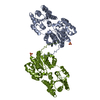
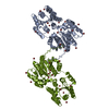
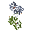
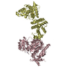
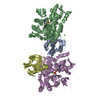
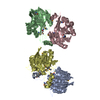
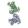
[English] 日本語
Yorodumi
- PDB-5wd7: Structure of a bacterial polysialyltransferase in complex with fo... -

+
Open data


ID or keywords:

Loading...

-
Basic information

Entry
Database: PDB / ID: 5wd7
TitleStructure of a bacterial polysialyltransferase in complex with fondaparinux
ComponentsSiaD
KeywordsMEMBRANE PROTEIN / polysialyltransferase / GT-B
Function / homologyAlpha-2,8-polysialyltransferase / Alpha-2,8-polysialyltransferase (POLYST) / nucleotide binding / fondaparinux / SiaD
Function and homology information
Biological speciesMannheimia haemolytica (bacteria)
MethodX-RAY DIFFRACTION / SYNCHROTRON / MOLECULAR REPLACEMENT / Resolution: 3.1 Å
AuthorsWorrall, L.J. / Lizak, C. / Strynadka, N.C.J.
CitationJournal: Sci Rep / Year: 2017
Title: X-ray crystallographic structure of a bacterial polysialyltransferase provides insight into the biosynthesis of capsular polysialic acid.
Authors: Lizak, C. / Worrall, L.J. / Baumann, L. / Pfleiderer, M.M. / Volkers, G. / Sun, T. / Sim, L. / Wakarchuk, W. / Withers, S.G. / Strynadka, N.C.J.
History
DepositionJul 4, 2017Deposition site: RCSB / Processing site: RCSB
Revision 1.0Aug 2, 2017Provider: repository / Type: Initial release
Revision 2.0Jul 29, 2020Group: Atomic model / Data collection ...Atomic model / Data collection / Derived calculations / Non-polymer description / Refinement description / Structure summary
Category: atom_site / chem_comp ...atom_site / chem_comp / entity / entity_name_com / pdbx_branch_scheme / pdbx_chem_comp_identifier / pdbx_entity_branch / pdbx_entity_branch_descriptor / pdbx_entity_branch_link / pdbx_entity_branch_list / pdbx_entity_nonpoly / pdbx_molecule_features / pdbx_nonpoly_scheme / refine_hist / struct_conn / struct_site / struct_site_gen
Item: _atom_site.B_iso_or_equiv / _atom_site.Cartn_x ..._atom_site.B_iso_or_equiv / _atom_site.Cartn_x / _atom_site.Cartn_y / _atom_site.Cartn_z / _atom_site.auth_asym_id / _atom_site.auth_atom_id / _atom_site.auth_comp_id / _atom_site.auth_seq_id / _atom_site.label_atom_id / _atom_site.label_comp_id / _atom_site.type_symbol / _chem_comp.formula / _chem_comp.formula_weight / _chem_comp.id / _chem_comp.mon_nstd_flag / _chem_comp.name / _chem_comp.pdbx_synonyms / _chem_comp.type / _entity.formula_weight / _entity.pdbx_description / _entity.src_method / _entity.type / _refine_hist.d_res_low
Description: Carbohydrate remediation / Provider: repository / Type: Remediation
Revision 2.1Mar 13, 2024Group: Data collection / Database references ...Data collection / Database references / Refinement description / Structure summary
Category: chem_comp / chem_comp_atom ...chem_comp / chem_comp_atom / chem_comp_bond / database_2 / struct_ncs_dom_lim
Item: _chem_comp.pdbx_synonyms / _database_2.pdbx_DOI ..._chem_comp.pdbx_synonyms / _database_2.pdbx_DOI / _database_2.pdbx_database_accession / _struct_ncs_dom_lim.beg_auth_comp_id / _struct_ncs_dom_lim.beg_label_asym_id / _struct_ncs_dom_lim.beg_label_comp_id / _struct_ncs_dom_lim.beg_label_seq_id / _struct_ncs_dom_lim.end_auth_comp_id / _struct_ncs_dom_lim.end_label_asym_id / _struct_ncs_dom_lim.end_label_comp_id / _struct_ncs_dom_lim.end_label_seq_id

-
Structure visualization

Structure viewerMolecule:
MolmilJmol/JSmol

Downloads & links

-
Assembly

Deposited unit
A: SiaD
M: SiaD
hetero molecules


Theoretical massNumber of molelcules
Total (without water)92,4315
Polymers90,7302
Non-polymers1,7003
Water00
1


  • Idetical with deposited unit
  • defined by author&software
  • Evidence: gel filtration
TypeNameSymmetry operationNumber
identity operation1_555x,y,z1
Buried area4120 Å2
ΔGint-40 kcal/mol
Surface area37320 Å2
MethodPISA
Unit cell
Length a, b, c (Å)78.312, 78.312, 299.895
Angle α, β, γ (deg.)90.00, 90.00, 120.00
Int Tables number152
Space group name H-MP3121
Noncrystallographic symmetry (NCS)NCS domain:
IDEns-IDDetails
11A
21M

NCS domain segments:

Component-ID: _ / Ens-ID: 1 / Beg auth comp-ID: MET / Beg label comp-ID: MET / End auth comp-ID: ILE / End label comp-ID: ILE / Refine code: _ / Auth seq-ID: 20 - 401 / Label seq-ID: 1 - 382

Dom-IDAuth asym-IDLabel asym-ID
1AA
2MB

-
Components

#1: Protein SiaD


Mass: 45365.168 Da / Num. of mol.: 2
Source method: isolated from a genetically manipulated source
Source: (gene. exp.) Mannheimia haemolytica (bacteria) / Gene: siaD / Production host: Escherichia coli (E. coli) / References: UniProt: G4RIN4
#2: Polysaccharide 2-deoxy-6-O-sulfo-2-(sulfoamino)-alpha-D-glucopyranose-(1-4)-beta-D-glucopyranuronic acid-(1-4)-2- ...2-deoxy-6-O-sulfo-2-(sulfoamino)-alpha-D-glucopyranose-(1-4)-beta-D-glucopyranuronic acid-(1-4)-2-deoxy-3,6-di-O-sulfo-2-(sulfoamino)-alpha-D-glucopyranose-(1-4)-2-O-sulfo-alpha-L-idopyranuronic acid-(1-4)-methyl 2-deoxy-6-O-sulfo-2-(sulfoamino)-alpha-D-glucopyranoside / fondaparinux


Type: oligosaccharide, Oligosaccharide / Class: Anticoagulant / Mass: 1508.264 Da / Num. of mol.: 1
Source method: isolated from a genetically manipulated source
Details: oligosaccharide / References: fondaparinux
DescriptorTypeProgram
WURCS=2.0/5,5,4/[a2122h-1a_1-5_1*OC_2*NSO/3=O/3=O_6*OSO/3=O/3=O][a2121A-1a_1-5_2*OSO/3=O/3=O][a2122h-1a_1-5_2*NSO/3=O/3=O_3*OSO/3=O/3=O_6*OSO/3=O/3=O][a2122A-1b_1-5][a2122h-1a_1-5_2*NSO/3=O/3=O_6*OSO/3=O/3=O]/1-2-3-4-5/a4-b1_b4-c1_c4-d1_d4-e1WURCSPDB2Glycan 1.1.0
[][methyl]{[(1+1)][a-D-GlcpNSO36SO3]{[(4+1)][a-L-IdopA2SO3]{[(4+1)][a-D-GlcpNSO33SO36SO3]{[(4+1)][b-D-GlcpA]{[(4+1)][a-D-GlcpNSO36SO3]{}}}}}}LINUCSPDB-CARE
#3: Chemical ChemComp-SO4 / SULFATE ION


Mass: 96.063 Da / Num. of mol.: 2 / Source method: obtained synthetically / Formula: SO4

-
Experimental details

-
Experiment

ExperimentMethod: X-RAY DIFFRACTION / Number of used crystals: 1

-
Sample preparation

CrystalDensity Matthews: 2.93 Å3/Da / Density % sol: 58 %
Crystal growTemperature: 296 K / Method: vapor diffusion, sitting drop
Details: 17% - 24% PEG3350 (v/v), 140 - 250 mM Mg2SO4 and 100 mM MES pH 7.2

-
Data collection

DiffractionMean temperature: 100 K
Diffraction sourceSource: SYNCHROTRON / Site: CLSI / Beamline: 08B1-1 / Wavelength: 1.00635 Å
DetectorType: ADSC QUANTUM 315r / Detector: CCD / Date: Oct 15, 2015
RadiationProtocol: SINGLE WAVELENGTH / Monochromatic (M) / Laue (L): M / Scattering type: x-ray
Radiation wavelengthWavelength: 1.00635 Å / Relative weight: 1
ReflectionResolution: 3.1→44.93 Å / Num. obs: 20333 / % possible obs: 100 % / Redundancy: 7.2 % / CC1/2: 0.998 / Rmerge(I) obs: 0.137 / Rpim(I) all: 0.054 / Rrim(I) all: 0.148 / Net I/σ(I): 14.7
Reflection shell

Diffraction-ID: 1

Resolution (Å)Redundancy (%)Rmerge(I) obsCC1/2Rpim(I) allRrim(I) all% possible all
3.1-3.317.41.3920.5860.5451.497100
8.77-44.936.10.02210.0090.02499.1

-
Processing

Software
NameVersionClassification
REFMAC5.8.0158refinement
Aimless0.5.29data scaling
PDB_EXTRACT3.22data extraction
xia2data reduction
PHASERphasing
RefinementMethod to determine structure: MOLECULAR REPLACEMENT / Resolution: 3.1→44.93 Å / Cor.coef. Fo:Fc: 0.954 / Cor.coef. Fo:Fc free: 0.924 / SU B: 22.514 / SU ML: 0.363 / Cross valid method: THROUGHOUT / ESU R Free: 0.436 / Stereochemistry target values: MAXIMUM LIKELIHOOD / Details: HYDROGENS HAVE BEEN ADDED IN THE RIDING POSITIONS
RfactorNum. reflection% reflectionSelection details
Rfree0.23837 938 4.6 %RANDOM
Rwork0.18725 ---
obs0.1896 19333 99.9 %-
Solvent computationIon probe radii: 0.8 Å / Shrinkage radii: 0.8 Å / VDW probe radii: 1.2 Å / Solvent model: MASK
Displacement parametersBiso mean: 88.079 Å2
Baniso -1Baniso -2Baniso -3
1--0.83 Å2-0.42 Å20 Å2
2---0.83 Å20 Å2
3---2.7 Å2
Refinement stepCycle: 1 / Resolution: 3.1→44.93 Å
ProteinNucleic acidLigandSolventTotal
Num. atoms6380 0 101 0 6481
Refine LS restraints
Refine-IDTypeDev idealDev ideal targetNumber
X-RAY DIFFRACTIONr_bond_refined_d0.0120.0196649
X-RAY DIFFRACTIONr_bond_other_d0.0020.026418
X-RAY DIFFRACTIONr_angle_refined_deg1.6031.9878939
X-RAY DIFFRACTIONr_angle_other_deg0.965314971
X-RAY DIFFRACTIONr_dihedral_angle_1_deg6.6025768
X-RAY DIFFRACTIONr_dihedral_angle_2_deg38.69124.061293
X-RAY DIFFRACTIONr_dihedral_angle_3_deg18.645151353
X-RAY DIFFRACTIONr_dihedral_angle_4_deg17.4641533
X-RAY DIFFRACTIONr_chiral_restr0.0870.2990
X-RAY DIFFRACTIONr_gen_planes_refined0.0080.026994
X-RAY DIFFRACTIONr_gen_planes_other0.0020.021335
X-RAY DIFFRACTIONr_nbd_refined
X-RAY DIFFRACTIONr_nbd_other
X-RAY DIFFRACTIONr_nbtor_refined
X-RAY DIFFRACTIONr_nbtor_other
X-RAY DIFFRACTIONr_xyhbond_nbd_refined
X-RAY DIFFRACTIONr_xyhbond_nbd_other
X-RAY DIFFRACTIONr_metal_ion_refined
X-RAY DIFFRACTIONr_metal_ion_other
X-RAY DIFFRACTIONr_symmetry_vdw_refined
X-RAY DIFFRACTIONr_symmetry_vdw_other
X-RAY DIFFRACTIONr_symmetry_hbond_refined
X-RAY DIFFRACTIONr_symmetry_hbond_other
X-RAY DIFFRACTIONr_symmetry_metal_ion_refined
X-RAY DIFFRACTIONr_symmetry_metal_ion_other
X-RAY DIFFRACTIONr_mcbond_it6.0028.5213069
X-RAY DIFFRACTIONr_mcbond_other6.0028.5213068
X-RAY DIFFRACTIONr_mcangle_it9.312.793838
X-RAY DIFFRACTIONr_mcangle_other9.29912.7893839
X-RAY DIFFRACTIONr_scbond_it6.3449.2283580
X-RAY DIFFRACTIONr_scbond_other6.3439.2283581
X-RAY DIFFRACTIONr_scangle_it
X-RAY DIFFRACTIONr_scangle_other9.99813.5595102
X-RAY DIFFRACTIONr_long_range_B_refined15.06526780
X-RAY DIFFRACTIONr_long_range_B_other15.06526781
X-RAY DIFFRACTIONr_rigid_bond_restr
X-RAY DIFFRACTIONr_sphericity_free
X-RAY DIFFRACTIONr_sphericity_bonded
Refine LS restraints NCS

Ens-ID: 1 / Number: 23666 / Refine-ID: X-RAY DIFFRACTION / Type: interatomic distance / Rms dev position: 0.17 Å / Weight position: 0.05

Dom-IDAuth asym-ID
1A
2M
LS refinement shellResolution: 3.1→3.181 Å / Total num. of bins used: 20
RfactorNum. reflection% reflection
Rfree0.358 66 -
Rwork0.338 1376 -
obs--99.86 %

+
About Yorodumi

-
News

-
Feb 9, 2022. New format data for meta-information of EMDB entries

New format data for meta-information of EMDB entries

  • Version 3 of the EMDB header file is now the official format.
  • The previous official version 1.9 will be removed from the archive.

Related info.:EMDB header

External links:wwPDB to switch to version 3 of the EMDB data model

-
Aug 12, 2020. Covid-19 info

Covid-19 info

URL: https://pdbj.org/emnavi/covid19.php

New page: Covid-19 featured information page in EM Navigator.

Related info.:Covid-19 info / Mar 5, 2020. Novel coronavirus structure data

+
Mar 5, 2020. Novel coronavirus structure data

Novel coronavirus structure data

Related info.:Yorodumi Speices / Aug 12, 2020. Covid-19 info

External links:COVID-19 featured content - PDBj / Molecule of the Month (242):Coronavirus Proteases

+
Jan 31, 2019. EMDB accession codes are about to change! (news from PDBe EMDB page)

EMDB accession codes are about to change! (news from PDBe EMDB page)

  • The allocation of 4 digits for EMDB accession codes will soon come to an end. Whilst these codes will remain in use, new EMDB accession codes will include an additional digit and will expand incrementally as the available range of codes is exhausted. The current 4-digit format prefixed with “EMD-” (i.e. EMD-XXXX) will advance to a 5-digit format (i.e. EMD-XXXXX), and so on. It is currently estimated that the 4-digit codes will be depleted around Spring 2019, at which point the 5-digit format will come into force.
  • The EM Navigator/Yorodumi systems omit the EMD- prefix.

Related info.:Q: What is EMD? / ID/Accession-code notation in Yorodumi/EM Navigator

External links:EMDB Accession Codes are Changing Soon! / Contact to PDBj

+
Jul 12, 2017. Major update of PDB

Major update of PDB

  • wwPDB released updated PDB data conforming to the new PDBx/mmCIF dictionary.
  • This is a major update changing the version number from 4 to 5, and with Remediation, in which all the entries are updated.
  • In this update, many items about electron microscopy experimental information are reorganized (e.g. em_software).
  • Now, EM Navigator and Yorodumi are based on the updated data.

External links:wwPDB Remediation / Enriched Model Files Conforming to OneDep Data Standards Now Available in the PDB FTP Archive

-
Yorodumi

Thousand views of thousand structures

  • Yorodumi is a browser for structure data from EMDB, PDB, SASBDB, etc.
  • This page is also the successor to EM Navigator detail page, and also detail information page/front-end page for Omokage search.
  • The word "yorodu" (or yorozu) is an old Japanese word meaning "ten thousand". "mi" (miru) is to see.

Related info.:EMDB / PDB / SASBDB / Comparison of 3 databanks / Yorodumi Search / Aug 31, 2016. New EM Navigator & Yorodumi / Yorodumi Papers / Jmol/JSmol / Function and homology information / Changes in new EM Navigator and Yorodumi

Read more