 Movie
Movie Controller

[English] 日本語
 Yorodumi
- PDB-5w6t: Crystal structure of the A/Puerto Rico/8/1934 (H1N1) influenza vi... -
+ Open data

- Basic information
| Entry | Database: PDB / ID: 5w6t | |||||||||||||||
|---|---|---|---|---|---|---|---|---|---|---|---|---|---|---|---|---|
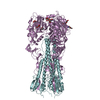
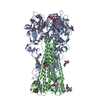
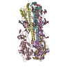
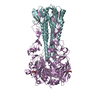
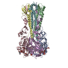
| Title | Crystal structure of the A/Puerto Rico/8/1934 (H1N1) influenza virus hemagglutinin in complex with cyclic peptide CP151070 (P7) | |||||||||||||||
 Components Components |
| |||||||||||||||
 Keywords Keywords | VIRAL PROTEIN/PEPTIDE / Glycoprotein / Ectodomain / N-glycosylation / VIRAL PROTEIN / VIRAL PROTEIN-PEPTIDE complex | |||||||||||||||
| Function / homology |  Function and homology information Function and homology informationTransport of HA trimer, NA tetramer and M2 tetramer from the endoplasmic reticulum to the Golgi Apparatus / Assembly of Viral Components at the Budding Site / Influenza Infection / Fusion of the Influenza Virion to the Host Cell Endosome / Release / Budding / Packaging of Eight RNA Segments / Uncoating of the Influenza Virion / Entry of Influenza Virion into Host Cell via Endocytosis / Viral mRNA Translation ...Transport of HA trimer, NA tetramer and M2 tetramer from the endoplasmic reticulum to the Golgi Apparatus / Assembly of Viral Components at the Budding Site / Influenza Infection / Fusion of the Influenza Virion to the Host Cell Endosome / Release / Budding / Packaging of Eight RNA Segments / Uncoating of the Influenza Virion / Entry of Influenza Virion into Host Cell via Endocytosis / Viral mRNA Translation / viral budding from plasma membrane / Immunoregulatory interactions between a Lymphoid and a non-Lymphoid cell / clathrin-dependent endocytosis of virus by host cell / host cell surface receptor binding / fusion of virus membrane with host plasma membrane / fusion of virus membrane with host endosome membrane / viral envelope / virion attachment to host cell / host cell plasma membrane / virion membrane / extracellular region / plasma membrane Similarity search - Function | |||||||||||||||
| Biological species |   Influenza A virus Influenza A virussynthetic construct (others) | |||||||||||||||
| Method |  X-RAY DIFFRACTION / SYNCHROTRON / MOLECULAR REPLACEMENT / Resolution: 2.59 Å X-RAY DIFFRACTION / SYNCHROTRON / MOLECULAR REPLACEMENT / Resolution: 2.59 Å | |||||||||||||||
 Authors Authors | Wilson, I.A. / Kadam, R.U. | |||||||||||||||
| Funding support |  United States, 1items United States, 1items
| |||||||||||||||
 Citation Citation | Journal: Science / Year: 2017 Title: Potent peptidic fusion inhibitors of influenza virus. Authors: Kadam, R.U. / Juraszek, J. / Brandenburg, B. / Buyck, C. / Schepens, W.B.G. / Kesteleyn, B. / Stoops, B. / Vreeken, R.J. / Vermond, J. / Goutier, W. / Tang, C. / Vogels, R. / Friesen, R.H.E. ...Authors: Kadam, R.U. / Juraszek, J. / Brandenburg, B. / Buyck, C. / Schepens, W.B.G. / Kesteleyn, B. / Stoops, B. / Vreeken, R.J. / Vermond, J. / Goutier, W. / Tang, C. / Vogels, R. / Friesen, R.H.E. / Goudsmit, J. / van Dongen, M.J.P. / Wilson, I.A. | |||||||||||||||
| History |
|
- Structure visualization
| Structure viewer | Molecule: MolmilJmol/JSmol |
|---|
- Downloads & links
-Download
| PDBx/mmCIF format | 5w6t.cif.gz | 122.7 KB | Display | PDBx/mmCIF format |
|---|---|---|---|---|
| PDB format | pdb5w6t.ent.gz | 92.1 KB | Display | PDB format |
| PDBx/mmJSON format | 5w6t.json.gz | Tree view | PDBx/mmJSON format | |
| Others |  Other downloads Other downloads |
-Validation report
| Arichive directory | https://data.pdbj.org/pub/pdb/validation_reports/w6/5w6tftp://data.pdbj.org/pub/pdb/validation_reports/w6/5w6t | HTTPS FTP |
|---|
-Related structure data
| Related structure data |  5w5sC  5w5uC  5w6iC  5w6rC  5w6uC  1ru7S C: citing same article ( S: Starting model for refinement |
|---|---|
| Similar structure data |


-Links
 PDBj
PDBj


- Assembly
| Deposited unit | 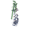
| |||||||||
|---|---|---|---|---|---|---|---|---|---|---|
| 1 | 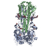
| |||||||||
| Unit cell |
| |||||||||
| Components on special symmetry positions |
|
-Components
-Protein , 2 types, 2 molecules AB
| #1: Protein | Mass: 36650.293 Da / Num. of mol.: 1 / Fragment: residues 18-343 Source method: isolated from a genetically manipulated source Source: (gene. exp.) Influenza A virus (strain A/Puerto Rico/8/1934 H1N1)Strain: A/Puerto Rico/8/1934 H1N1 / Gene: HA / Production host:  Trichoplusia ni (cabbage looper) / References: UniProt: P03452 Trichoplusia ni (cabbage looper) / References: UniProt: P03452 |
|---|---|
| #2: Protein | Mass: 20138.393 Da / Num. of mol.: 1 / Fragment: residues 344-519 Source method: isolated from a genetically manipulated source Source: (gene. exp.) Influenza A virus (strain A/Puerto Rico/8/1934 H1N1)Strain: A/Puerto Rico/8/1934 H1N1 / Gene: HA / Production host: Trichoplusia ni (cabbage looper) / References: UniProt: P03452 |
-Protein/peptide , 1 types, 1 molecules F
| #3: Protein/peptide | Mass: 1641.691 Da / Num. of mol.: 1 / Source method: obtained synthetically / Source: (synth.) synthetic construct (others) |
|---|
-Sugars , 2 types, 3 molecules 
| #4: Polysaccharide | 2-acetamido-2-deoxy-beta-D-glucopyranose-(1-4)-2-acetamido-2-deoxy-beta-D-glucopyranose Source method: isolated from a genetically manipulated source |
|---|---|
| #5: Sugar |  Type: D-saccharide, beta linking / Mass: 221.208 Da / Num. of mol.: 2 Type: D-saccharide, beta linking / Mass: 221.208 Da / Num. of mol.: 2Source method: isolated from a genetically manipulated source Formula: C8H15NO6 |
-Non-polymers , 3 types, 71 molecules 


| #6: Chemical |  Mass: 92.094 Da / Num. of mol.: 2 / Source method: obtained synthetically / Formula: C3H8O3 Mass: 92.094 Da / Num. of mol.: 2 / Source method: obtained synthetically / Formula: C3H8O3#7: Chemical | ChemComp-CL / |  Mass: 35.453 Da / Num. of mol.: 1 / Source method: obtained synthetically / Formula: Cl Mass: 35.453 Da / Num. of mol.: 1 / Source method: obtained synthetically / Formula: Cl#8: Water | ChemComp-HOH / | Mass: 18.015 Da / Num. of mol.: 68 / Source method: isolated from a natural source / Formula: H2O |
|---|
-Details
| Has protein modification | Y |
|---|
-Experimental details
-Experiment
| Experiment | Method: X-RAY DIFFRACTION / Number of used crystals: 1 |
|---|
- Sample preparation
| Crystal | Density Matthews: 3.26 Å3/Da / Density % sol: 62.22 % |
|---|---|
| Crystal grow | Temperature: 277 K / Method: vapor diffusion, sitting drop / pH: 5.3 Details: HA: 10 mg/ml 1M lithium chloride 20 % w/v PEG 6000 0.1M MES, pH=5.3 |
-Data collection
| Diffraction | Mean temperature: 80 K |
|---|---|
| Diffraction source | Source: SYNCHROTRON / Site: SSRL / Beamline: BL12-2 / Wavelength: 1.0331 Å |
| Detector | Type: DECTRIS PILATUS 6M / Detector: PIXEL / Date: Dec 14, 2015 |
| Radiation | Protocol: SINGLE WAVELENGTH / Monochromatic (M) / Laue (L): M / Scattering type: x-ray |
| Radiation wavelength | Wavelength: 1.0331 Å / Relative weight: 1 |
| Reflection | Resolution: 2.59→50 Å / Num. obs: 21313 / % possible obs: 92.2 % / Redundancy: 3.1 % / Rsym value: 0.12 / Net I/σ(I): 14.8 |
| Reflection shell | Resolution: 2.59→2.63 Å / Redundancy: 3 % / Mean I/σ(I) obs: 1.2 / Rsym value: 0.8 / % possible all: 90.9 |
- Processing
| Software |
| |||||||||||||||||||||||||||||||||||||||||||||||||||||||||||||||
|---|---|---|---|---|---|---|---|---|---|---|---|---|---|---|---|---|---|---|---|---|---|---|---|---|---|---|---|---|---|---|---|---|---|---|---|---|---|---|---|---|---|---|---|---|---|---|---|---|---|---|---|---|---|---|---|---|---|---|---|---|---|---|---|---|
| Refinement | Method to determine structure: MOLECULAR REPLACEMENT Starting model: 1RU7 Resolution: 2.59→41.08 Å / SU ML: 0.4 / Cross valid method: FREE R-VALUE / σ(F): 1.36 / Phase error: 28.02
| |||||||||||||||||||||||||||||||||||||||||||||||||||||||||||||||
| Solvent computation | Shrinkage radii: 0.9 Å / VDW probe radii: 1.11 Å | |||||||||||||||||||||||||||||||||||||||||||||||||||||||||||||||
| Refinement step | Cycle: LAST / Resolution: 2.59→41.08 Å
| |||||||||||||||||||||||||||||||||||||||||||||||||||||||||||||||
| Refine LS restraints |
| |||||||||||||||||||||||||||||||||||||||||||||||||||||||||||||||
| LS refinement shell |
|
