 Movie
Movie Controller

[English] 日本語
 Yorodumi
- PDB-5w3e: CryoEM structure of rhinovirus B14 in complex with C5 Fab (33 deg... -
+ Open data

- Basic information
| Entry | Database: PDB / ID: 5w3e | ||||||
|---|---|---|---|---|---|---|---|
| Title | CryoEM structure of rhinovirus B14 in complex with C5 Fab (33 degrees Celsius, molar ratio 1:3, full particle) | ||||||
 Components Components |
| ||||||
 Keywords Keywords | VIRUS/IMMUNE SYSTEM / virus / antibody / VIRUS-IMMUNE SYSTEM complex | ||||||
| Function / homology |  Function and homology information Function and homology informationlysis of host organelle involved in viral entry into host cell / symbiont-mediated suppression of host cytoplasmic pattern recognition receptor signaling pathway via inhibition of RIG-I activity / picornain 2A / symbiont-mediated suppression of host mRNA export from nucleus / symbiont genome entry into host cell via pore formation in plasma membrane / picornain 3C / T=pseudo3 icosahedral viral capsid / host cell cytoplasmic vesicle membrane / nucleoside-triphosphate phosphatase / channel activity ...lysis of host organelle involved in viral entry into host cell / symbiont-mediated suppression of host cytoplasmic pattern recognition receptor signaling pathway via inhibition of RIG-I activity / picornain 2A / symbiont-mediated suppression of host mRNA export from nucleus / symbiont genome entry into host cell via pore formation in plasma membrane / picornain 3C / T=pseudo3 icosahedral viral capsid / host cell cytoplasmic vesicle membrane / nucleoside-triphosphate phosphatase / channel activity / monoatomic ion transmembrane transport / DNA replication / RNA helicase activity / endocytosis involved in viral entry into host cell / symbiont-mediated activation of host autophagy / RNA-directed RNA polymerase / cysteine-type endopeptidase activity / viral RNA genome replication / RNA-directed RNA polymerase activity / DNA-templated transcription / virion attachment to host cell / host cell nucleus / structural molecule activity / ATP hydrolysis activity / proteolysis / RNA binding / zinc ion binding / ATP binding / membrane Similarity search - Function | ||||||
| Biological species |   Human rhinovirus 14 Human rhinovirus 14 | ||||||
| Method | ELECTRON MICROSCOPY / single particle reconstruction / cryo EM / Resolution: 2.53 Å | ||||||
 Authors Authors | Liu, Y. / Dong, Y. / Rossmann, M.G. | ||||||
| Funding support |  United States, 1items United States, 1items
| ||||||
 Citation Citation | Journal: Proc Natl Acad Sci U S A / Year: 2017 Title: Antibody-induced uncoating of human rhinovirus B14. Authors: Yangchao Dong / Yue Liu / Wen Jiang / Thomas J Smith / Zhikai Xu / Michael G Rossmann /  Abstract: Rhinoviruses (RVs) are the major causes of common colds in humans. They have a nonenveloped, icosahedral capsid surrounding a positive-strand RNA genome. Here we report that the antigen-binding (Fab) ...Rhinoviruses (RVs) are the major causes of common colds in humans. They have a nonenveloped, icosahedral capsid surrounding a positive-strand RNA genome. Here we report that the antigen-binding (Fab) fragment of a neutralizing antibody (C5) can trigger genome release from RV-B14 to form emptied particles and neutralize virus infection. Using cryo-electron microscopy, structures of the C5 Fab in complex with the full and emptied particles have been determined at 2.3 Å and 3.0 Å resolution, respectively. Each of the 60 Fab molecules binds primarily to a region on viral protein 3 (VP3). Binding of the C5 Fabs to RV-B14 results in significant conformational changes around holes in the capsid through which the viral RNA might exit. These results are so far the highest resolution view of an antibody-virus complex and elucidate a mechanism whereby antibodies neutralize RVs and related viruses by inducing virus uncoating. | ||||||
| History |
|
- Structure visualization
| Movie |
Movie viewer |
|---|---|
| Structure viewer | Molecule: MolmilJmol/JSmol |
- Downloads & links
-Download
| PDBx/mmCIF format | 5w3e.cif.gz | 214.5 KB | Display | PDBx/mmCIF format |
|---|---|---|---|---|
| PDB format | pdb5w3e.ent.gz | 168 KB | Display | PDB format |
| PDBx/mmJSON format | 5w3e.json.gz | Tree view | PDBx/mmJSON format | |
| Others |  Other downloads Other downloads |
-Validation report
| Summary document | 5w3e_validation.pdf.gz | 1.1 MB | Display | wwPDB validaton report |
|---|---|---|---|---|
| Full document | 5w3e_full_validation.pdf.gz | 1.1 MB | Display | |
| Data in XML | 5w3e_validation.xml.gz | 55.9 KB | Display | |
| Data in CIF | 5w3e_validation.cif.gz | 84.3 KB | Display | |
| Arichive directory | https://data.pdbj.org/pub/pdb/validation_reports/w3/5w3eftp://data.pdbj.org/pub/pdb/validation_reports/w3/5w3e | HTTPS FTP |
-Related structure data
| Related structure data | 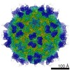 8754MC 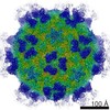 8761C 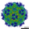 8762C 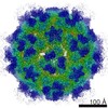 8763C 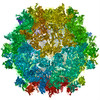 5w3lC 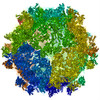 5w3mC 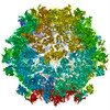 5w3oC M: map data used to model this data C: citing same article ( |
|---|---|
| Similar structure data |


-Links
 PDBj
PDBj


- Assembly
| Deposited unit | 
|
|---|---|
| 1 | x 60
|
| 2 |
|
| 3 | x 5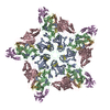
|
| 4 | x 6
|
| 5 | 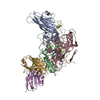
|
| Symmetry | Point symmetry: (Schoenflies symbol: I (icosahedral)) |
-Components
-Viral protein ... , 4 types, 4 molecules ABCD
| #3: Protein | Mass: 32560.549 Da / Num. of mol.: 1 / Fragment: UNP residues 568-856 / Source method: isolated from a natural source / Source: (natural) Human rhinovirus 14 / References: UniProt: P03303 |
|---|---|
| #4: Protein | Mass: 26236.754 Da / Num. of mol.: 1 / Fragment: UNP residues 332-567 / Source method: isolated from a natural source / Source: (natural) Human rhinovirus 14 / References: UniProt: P03303 |
| #5: Protein | Mass: 28501.361 Da / Num. of mol.: 1 / Fragment: UNP residues 70-331 / Source method: isolated from a natural source / Source: (natural) Human rhinovirus 14 / References: UniProt: P03303 |
| #6: Protein | Mass: 7183.863 Da / Num. of mol.: 1 / Fragment: UNP residues 2-69 / Source method: isolated from a natural source / Source: (natural) Human rhinovirus 14 / References: UniProt: P03303 |
-Antibody , 2 types, 2 molecules EG
| #1: Antibody | Mass: 11989.335 Da / Num. of mol.: 1 / Source method: isolated from a natural source / Source: (natural) |
|---|---|
| #2: Antibody | Mass: 10897.161 Da / Num. of mol.: 1 / Source method: isolated from a natural source / Source: (natural) |
-Non-polymers , 1 types, 216 molecules 
| #7: Water | ChemComp-HOH / Mass: 18.015 Da / Num. of mol.: 216 / Source method: isolated from a natural source / Formula: H2O |
|---|
-Details
| Has protein modification | Y |
|---|
-Experimental details
-Experiment
| Experiment | Method: ELECTRON MICROSCOPY |
|---|---|
| EM experiment | Aggregation state: PARTICLE / 3D reconstruction method: single particle reconstruction |
- Sample preparation
| Component | Name: Human rhinovirus B14 / Type: VIRUS / Details: Viruses were grown in HeLa-H1 cells. / Entity ID: #1-#6 / Source: NATURAL |
|---|---|
| Source (natural) | Organism: Human rhinovirus B14 |
| Details of virus | Empty: NO / Enveloped: NO / Isolate: STRAIN / Type: VIRION |
| Buffer solution | pH: 8 Details: 20 mM Tris, 120 mM sodium chloride, 1 mM EDTA, pH 8.0 |
| Specimen | Embedding applied: NO / Shadowing applied: NO / Staining applied: NO / Vitrification applied: YES |
| Specimen support | Grid material: COPPER / Grid mesh size: 400 divisions/in. / Grid type: Ted Pella, Ultrathin lacey carbon |
| Vitrification | Instrument: GATAN CRYOPLUNGE 3 / Cryogen name: ETHANE / Humidity: 85 % / Chamber temperature: 298 K |
- Electron microscopy imaging
| Experimental equipment |  Model: Titan Krios / Image courtesy: FEI Company |
|---|---|
| Microscopy | Model: FEI TITAN KRIOS |
| Electron gun | Electron source:  FIELD EMISSION GUN / Accelerating voltage: 300 kV / Illumination mode: FLOOD BEAM FIELD EMISSION GUN / Accelerating voltage: 300 kV / Illumination mode: FLOOD BEAM |
| Electron lens | Mode: BRIGHT FIELD / Nominal magnification: 29000 X / Nominal defocus max: 3000 nm / Nominal defocus min: 1000 nm / Cs: 2.7 mm / C2 aperture diameter: 100 µm / Alignment procedure: COMA FREE |
| Specimen holder | Cryogen: NITROGEN / Specimen holder model: FEI TITAN KRIOS AUTOGRID HOLDER |
| Image recording | Average exposure time: 6 sec. / Electron dose: 30 e/Å2 / Detector mode: COUNTING / Film or detector model: GATAN K2 SUMMIT (4k x 4k) / Num. of grids imaged: 1 / Num. of real images: 1111 |
| Image scans | Sampling size: 5 µm / Width: 3710 / Height: 3838 / Movie frames/image: 50 / Used frames/image: 4-49 |
- Processing
| EM software |
| ||||||||||||||||||||||||||||||||||||||||||||||||
|---|---|---|---|---|---|---|---|---|---|---|---|---|---|---|---|---|---|---|---|---|---|---|---|---|---|---|---|---|---|---|---|---|---|---|---|---|---|---|---|---|---|---|---|---|---|---|---|---|---|
| CTF correction | Details: On-the-fly CTF correction during 2D alignment and 3D reconstruction Type: PHASE FLIPPING AND AMPLITUDE CORRECTION | ||||||||||||||||||||||||||||||||||||||||||||||||
| Particle selection | Num. of particles selected: 60065 | ||||||||||||||||||||||||||||||||||||||||||||||||
| Symmetry | Point symmetry: I (icosahedral) | ||||||||||||||||||||||||||||||||||||||||||||||||
| 3D reconstruction | Resolution: 2.53 Å / Resolution method: FSC 0.143 CUT-OFF / Num. of particles: 23242 / Algorithm: FOURIER SPACE / Symmetry type: POINT | ||||||||||||||||||||||||||||||||||||||||||||||||
| Atomic model building | Protocol: RIGID BODY FIT / Space: REAL / Target criteria: Correlation coeffcient Details: A combination of the following approaches was used: (1) model rebuilding using Coot, (2) real space refinement using Phenix, (3) reciprocal space refinment using Phenix (as in standard ...Details: A combination of the following approaches was used: (1) model rebuilding using Coot, (2) real space refinement using Phenix, (3) reciprocal space refinment using Phenix (as in standard crystallographic refinement). |
