 Movie
Movie Controller

[English] 日本語
 Yorodumi
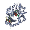
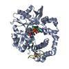
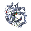
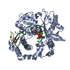
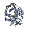
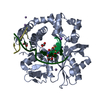
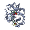
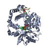
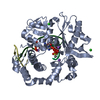
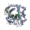
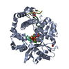
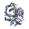
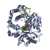
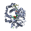
- PDB-5vzb: Post-catalytic complex of human Polymerase Mu (G433S) mutant with... -
+ Open data

- Basic information
| Entry | Database: PDB / ID: 5vzb | |||||||||||||||
|---|---|---|---|---|---|---|---|---|---|---|---|---|---|---|---|---|
| Title | Post-catalytic complex of human Polymerase Mu (G433S) mutant with incoming UTP | |||||||||||||||
 Components Components |
| |||||||||||||||
 Keywords Keywords | TRANSFERASE/DNA / Family X / nonhomologous end-joining / DNA double strand break repair / ribonucleotide incorporation / TRANSFERASE-DNA complex | |||||||||||||||
| Function / homology |  Function and homology information Function and homology informationsomatic hypermutation of immunoglobulin genes / B cell differentiation / Nonhomologous End-Joining (NHEJ) / double-strand break repair via nonhomologous end joining / DNA recombination / DNA-directed DNA polymerase / DNA-directed DNA polymerase activity / DNA binding / nucleoplasm / metal ion binding / nucleus Similarity search - Function | |||||||||||||||
| Biological species |  Homo sapiens (human) Homo sapiens (human)synthetic construct (others) | |||||||||||||||
| Method |  X-RAY DIFFRACTION / SYNCHROTRON / MOLECULAR REPLACEMENT / Resolution: 1.5 Å X-RAY DIFFRACTION / SYNCHROTRON / MOLECULAR REPLACEMENT / Resolution: 1.5 Å | |||||||||||||||
 Authors Authors | Moon, A.F. / Pryor, J.M. / Ramsden, D.A. / Kunkel, T.A. / Bebenek, K. / Pedersen, L.C. | |||||||||||||||
| Funding support |  United States, 4items United States, 4items
| |||||||||||||||
 Citation Citation | Journal: Nucleic Acids Res. / Year: 2017 Title: Structural accommodation of ribonucleotide incorporation by the DNA repair enzyme polymerase Mu. Authors: Moon, A.F. / Pryor, J.M. / Ramsden, D.A. / Kunkel, T.A. / Bebenek, K. / Pedersen, L.C. | |||||||||||||||
| History |
|
- Structure visualization
| Structure viewer | Molecule: MolmilJmol/JSmol |
|---|
- Downloads & links
-Download
| PDBx/mmCIF format | 5vzb.cif.gz | 186.8 KB | Display | PDBx/mmCIF format |
|---|---|---|---|---|
| PDB format | pdb5vzb.ent.gz | 140 KB | Display | PDB format |
| PDBx/mmJSON format | 5vzb.json.gz | Tree view | PDBx/mmJSON format | |
| Others |  Other downloads Other downloads |
-Validation report
| Arichive directory | https://data.pdbj.org/pub/pdb/validation_reports/vz/5vzbftp://data.pdbj.org/pub/pdb/validation_reports/vz/5vzb | HTTPS FTP |
|---|
-Related structure data
| Related structure data |  5twpC 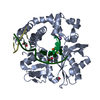 5twqC  5twrC 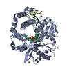 5twsC 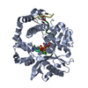 5vz7C  5vz8C 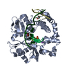 5vz9C 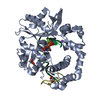 5vzaC 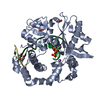 5vzcC  5vzdC  5vzeC 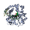 5vzfC  5vzgC 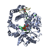 5vzhC  5vziC  4m04S C: citing same article ( S: Starting model for refinement |
|---|---|
| Similar structure data |


-Links
 PDBj
PDBj


- Assembly
| Deposited unit | 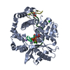
| ||||||||
|---|---|---|---|---|---|---|---|---|---|
| 1 |
| ||||||||
| Unit cell |
|
-Components
-DNA chain , 2 types, 2 molecules TD
| #2: DNA chain | Mass: 2740.812 Da / Num. of mol.: 1 / Source method: obtained synthetically / Source: (synth.) synthetic construct (others) |
|---|---|
| #4: DNA chain | Mass: 1191.818 Da / Num. of mol.: 1 / Source method: obtained synthetically / Source: (synth.) synthetic construct (others) |
-Protein / DNA/RNA hybrid , 2 types, 2 molecules AP
| #1: Protein | Mass: 39874.188 Da / Num. of mol.: 1 / Fragment: residues 134-494 / Mutation: G433S Source method: isolated from a genetically manipulated source Source: (gene. exp.) Homo sapiens (human) / Gene: POLM, polmu / Plasmid: pGEXM / Production host:  |
|---|---|
| #3: DNA/RNA hybrid | Mass: 1496.996 Da / Num. of mol.: 1 / Source method: obtained synthetically / Source: (synth.) synthetic construct (others) |
-Non-polymers , 6 types, 387 molecules 


| #5: Chemical |  Mass: 24.305 Da / Num. of mol.: 3 / Source method: obtained synthetically / Formula: Mg Mass: 24.305 Da / Num. of mol.: 3 / Source method: obtained synthetically / Formula: Mg#6: Chemical | ChemComp-NA / |  Mass: 22.990 Da / Num. of mol.: 1 / Source method: obtained synthetically / Formula: Na Mass: 22.990 Da / Num. of mol.: 1 / Source method: obtained synthetically / Formula: Na#7: Chemical |  Mass: 35.453 Da / Num. of mol.: 2 / Source method: obtained synthetically / Formula: Cl Mass: 35.453 Da / Num. of mol.: 2 / Source method: obtained synthetically / Formula: Cl#8: Chemical | ChemComp-UTP / |  Mass: 484.141 Da / Num. of mol.: 1 / Source method: obtained synthetically / Formula: C9H15N2O15P3 / Comment: UTP*YM Mass: 484.141 Da / Num. of mol.: 1 / Source method: obtained synthetically / Formula: C9H15N2O15P3 / Comment: UTP*YM#9: Chemical |  Mass: 62.068 Da / Num. of mol.: 2 / Source method: obtained synthetically / Formula: C2H6O2 Mass: 62.068 Da / Num. of mol.: 2 / Source method: obtained synthetically / Formula: C2H6O2#10: Water | ChemComp-HOH / | Mass: 18.015 Da / Num. of mol.: 378 / Source method: isolated from a natural source / Formula: H2O |
|---|
-Experimental details
-Experiment
| Experiment | Method: X-RAY DIFFRACTION / Number of used crystals: 1 |
|---|
- Sample preparation
| Crystal | Density Matthews: 2.5 Å3/Da / Density % sol: 50.72 % |
|---|---|
| Crystal grow | Temperature: 293 K / Method: vapor diffusion, sitting drop / pH: 7.5 / Details: 90mM HEPES pH 7.5, 18% PEG4K |
-Data collection
| Diffraction | Mean temperature: 100 K |
|---|---|
| Diffraction source | Source: SYNCHROTRON / Site: APS / Beamline: 22-ID / Wavelength: 1 Å |
| Detector | Type: MARMOSAIC 300 mm CCD / Detector: CCD / Date: Apr 13, 2017 / Details: Rosenbaum-Rock vertical focusing mirror |
| Radiation | Monochromator: Rosenbaum-Rock monochromator high-resolution double-crystal Si(220) sagittal focusing Protocol: SINGLE WAVELENGTH / Monochromatic (M) / Laue (L): M / Scattering type: x-ray |
| Radiation wavelength | Wavelength: 1 Å / Relative weight: 1 |
| Reflection | Resolution: 1.5→50 Å / Num. obs: 72904 / % possible obs: 99.5 % / Redundancy: 6.8 % / Rpim(I) all: 0.027 / Rsym value: 0.065 / Χ2: 0.933 / Net I/σ(I): 25.34 |
| Reflection shell | Resolution: 1.5→1.53 Å / Redundancy: 3.5 % / Mean I/σ(I) obs: 2.45 / Num. unique obs: 3437 / CC1/2: 0.688 / Rpim(I) all: 0.339 / Rsym value: 0.578 / Χ2: 0.836 / % possible all: 95.8 |
- Processing
| Software |
| ||||||||||||||||||||||||||||||||||||||||||||||||||||||||||||||||||||||||||||||||||||||||||||||||||||||||||||||||||||||||||||||||||||||||||||||||||||||||||||||||||||||||||||||||||||||
|---|---|---|---|---|---|---|---|---|---|---|---|---|---|---|---|---|---|---|---|---|---|---|---|---|---|---|---|---|---|---|---|---|---|---|---|---|---|---|---|---|---|---|---|---|---|---|---|---|---|---|---|---|---|---|---|---|---|---|---|---|---|---|---|---|---|---|---|---|---|---|---|---|---|---|---|---|---|---|---|---|---|---|---|---|---|---|---|---|---|---|---|---|---|---|---|---|---|---|---|---|---|---|---|---|---|---|---|---|---|---|---|---|---|---|---|---|---|---|---|---|---|---|---|---|---|---|---|---|---|---|---|---|---|---|---|---|---|---|---|---|---|---|---|---|---|---|---|---|---|---|---|---|---|---|---|---|---|---|---|---|---|---|---|---|---|---|---|---|---|---|---|---|---|---|---|---|---|---|---|---|---|---|---|
| Refinement | Method to determine structure: MOLECULAR REPLACEMENT Starting model: 4M04 Resolution: 1.5→32.328 Å / SU ML: 0.13 / Cross valid method: FREE R-VALUE / σ(F): 1.49 / Phase error: 17.2 Details: THE AUTHORS STATE THAT THE ANOMALOUS SIGNAL IS OBSERVED AT ONE POSITION IN THIS STRUCTURE--AT THE METAL OCCUPYING THE HHH2 SITE (PEAK > 10 SIGMA). NO KNOWN ANOMALOUS SCATTERERS WERE ADDED, ...Details: THE AUTHORS STATE THAT THE ANOMALOUS SIGNAL IS OBSERVED AT ONE POSITION IN THIS STRUCTURE--AT THE METAL OCCUPYING THE HHH2 SITE (PEAK > 10 SIGMA). NO KNOWN ANOMALOUS SCATTERERS WERE ADDED, SO THIS POSITION HAS BEEN PUTATIVELY MODELED AS SODIUM, WHICH IS CONSISTENT WITH ITS IDENTITY IN OTHER REPORTED STRUCTURES.
| ||||||||||||||||||||||||||||||||||||||||||||||||||||||||||||||||||||||||||||||||||||||||||||||||||||||||||||||||||||||||||||||||||||||||||||||||||||||||||||||||||||||||||||||||||||||
| Solvent computation | Shrinkage radii: 0.9 Å / VDW probe radii: 1.11 Å | ||||||||||||||||||||||||||||||||||||||||||||||||||||||||||||||||||||||||||||||||||||||||||||||||||||||||||||||||||||||||||||||||||||||||||||||||||||||||||||||||||||||||||||||||||||||
| Refinement step | Cycle: LAST / Resolution: 1.5→32.328 Å
| ||||||||||||||||||||||||||||||||||||||||||||||||||||||||||||||||||||||||||||||||||||||||||||||||||||||||||||||||||||||||||||||||||||||||||||||||||||||||||||||||||||||||||||||||||||||
| Refine LS restraints |
| ||||||||||||||||||||||||||||||||||||||||||||||||||||||||||||||||||||||||||||||||||||||||||||||||||||||||||||||||||||||||||||||||||||||||||||||||||||||||||||||||||||||||||||||||||||||
| LS refinement shell |
| ||||||||||||||||||||||||||||||||||||||||||||||||||||||||||||||||||||||||||||||||||||||||||||||||||||||||||||||||||||||||||||||||||||||||||||||||||||||||||||||||||||||||||||||||||||||
| Refinement TLS params. | Method: refined / Refine-ID: X-RAY DIFFRACTION
| ||||||||||||||||||||||||||||||||||||||||||||||||||||||||||||||||||||||||||||||||||||||||||||||||||||||||||||||||||||||||||||||||||||||||||||||||||||||||||||||||||||||||||||||||||||||
| Refinement TLS group |
|
