 Movie
Movie Controller


+ Open data

- Basic information
| Entry | Database: PDB / ID: 4m04 | ||||||
|---|---|---|---|---|---|---|---|
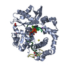
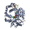
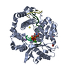
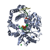
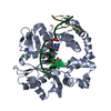
| Title | Human DNA Polymerase Mu ternary complex | ||||||
 Components Components |
| ||||||
 Keywords Keywords | TRANSFERASE/DNA / polymerase / DNA break repair / TRANSFERASE-DNA complex | ||||||
| Function / homology |  Function and homology information Function and homology informationsomatic hypermutation of immunoglobulin genes / B cell differentiation / Nonhomologous End-Joining (NHEJ) / double-strand break repair via nonhomologous end joining / DNA recombination / DNA-directed DNA polymerase / DNA-directed DNA polymerase activity / DNA binding / nucleoplasm / metal ion binding / nucleus Similarity search - Function | ||||||
| Biological species |  Homo sapiens (human) Homo sapiens (human) | ||||||
| Method |  X-RAY DIFFRACTION / MOLECULAR REPLACEMENT / molecular replacement / Resolution: 1.898 Å X-RAY DIFFRACTION / MOLECULAR REPLACEMENT / molecular replacement / Resolution: 1.898 Å | ||||||
 Authors Authors | Moon, A.F. / Pryor, J.M. / Ramsden, D.A. / Kunkel, T.A. / Bebenek, K. / Pedersen, L.C. | ||||||
 Citation Citation | Journal: Nat.Struct.Mol.Biol. / Year: 2014 Title: Sustained active site rigidity during synthesis by human DNA polymerase mu. Authors: Moon, A.F. / Pryor, J.M. / Ramsden, D.A. / Kunkel, T.A. / Bebenek, K. / Pedersen, L.C. | ||||||
| History |
|
- Structure visualization
| Structure viewer | Molecule: MolmilJmol/JSmol |
|---|
- Downloads & links
-Download
| PDBx/mmCIF format | 4m04.cif.gz | 110 KB | Display | PDBx/mmCIF format |
|---|---|---|---|---|
| PDB format | pdb4m04.ent.gz | 76.4 KB | Display | PDB format |
| PDBx/mmJSON format | 4m04.json.gz | Tree view | PDBx/mmJSON format | |
| Others |  Other downloads Other downloads |
-Validation report
| Arichive directory | https://data.pdbj.org/pub/pdb/validation_reports/m0/4m04ftp://data.pdbj.org/pub/pdb/validation_reports/m0/4m04 | HTTPS FTP |
|---|
-Related structure data
| Related structure data |  4lzdC  4lzgC  4m0aC  2ihmS S: Starting model for refinement C: citing same article ( |
|---|---|
| Similar structure data |


-Links
 PDBj
PDBj


- Assembly
| Deposited unit | 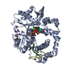
| ||||||||
|---|---|---|---|---|---|---|---|---|---|
| 1 |
| ||||||||
| Unit cell |
|
-Components
-Protein , 1 types, 1 molecules A
| #1: Protein | Mass: 40054.434 Da / Num. of mol.: 1 Fragment: Polymerase Mu Loop2 deletion variant, UNP residues 132-494 Source method: isolated from a genetically manipulated source Details: Modified version of the pGEX4T3 vector backbone, where a TEV protease cleavage site has been inserted downstream of the thrombin cleavage site, and the sequence of the multicloning site has ...Details: Modified version of the pGEX4T3 vector backbone, where a TEV protease cleavage site has been inserted downstream of the thrombin cleavage site, and the sequence of the multicloning site has been replaced by that from the pMALX vector (Moon et al, 2010). Three TGA stop codons have been inserted downstream of the MCS, each utilizing a different reading frame. Source: (gene. exp.) Homo sapiens (human) / Gene: POLM, polmu / Plasmid: pGEXM / Production host:  |
|---|
-DNA chain , 3 types, 3 molecules TPD
| #2: DNA chain | Mass: 2740.812 Da / Num. of mol.: 1 / Source method: obtained synthetically / Details: synthesized by Oligos Etc |
|---|---|
| #3: DNA chain | Mass: 1190.830 Da / Num. of mol.: 1 / Source method: obtained synthetically / Details: synthesized by Oligos Etc |
| #4: DNA chain | Mass: 1191.818 Da / Num. of mol.: 1 / Source method: obtained synthetically / Details: synthesized by Oligos Etc |
-Non-polymers , 7 types, 390 molecules 


| #5: Chemical | ChemComp-DUP /  Mass: 467.157 Da / Num. of mol.: 1 / Source method: obtained synthetically / Formula: C9H16N3O13P3 Mass: 467.157 Da / Num. of mol.: 1 / Source method: obtained synthetically / Formula: C9H16N3O13P3 | ||||||||||
|---|---|---|---|---|---|---|---|---|---|---|---|
| #6: Chemical |  Mass: 24.305 Da / Num. of mol.: 2 / Source method: obtained synthetically / Formula: Mg Mass: 24.305 Da / Num. of mol.: 2 / Source method: obtained synthetically / Formula: Mg#7: Chemical |  Mass: 62.068 Da / Num. of mol.: 3 / Source method: obtained synthetically / Formula: C2H6O2 Mass: 62.068 Da / Num. of mol.: 3 / Source method: obtained synthetically / Formula: C2H6O2#8: Chemical | ChemComp-CL / |  Mass: 35.453 Da / Num. of mol.: 1 / Source method: obtained synthetically / Formula: Cl Mass: 35.453 Da / Num. of mol.: 1 / Source method: obtained synthetically / Formula: Cl#9: Chemical | ChemComp-NA / |  Mass: 22.990 Da / Num. of mol.: 1 / Source method: obtained synthetically / Formula: Na Mass: 22.990 Da / Num. of mol.: 1 / Source method: obtained synthetically / Formula: Na#10: Chemical | ChemComp-EPE / |  Mass: 238.305 Da / Num. of mol.: 1 / Source method: obtained synthetically / Formula: C8H18N2O4S / Comment: pH buffer*YM Mass: 238.305 Da / Num. of mol.: 1 / Source method: obtained synthetically / Formula: C8H18N2O4S / Comment: pH buffer*YM#11: Water | ChemComp-HOH / | Mass: 18.015 Da / Num. of mol.: 381 / Source method: isolated from a natural source / Formula: H2O |
-Experimental details
-Experiment
| Experiment | Method: X-RAY DIFFRACTION / Number of used crystals: 1 |
|---|
- Sample preparation
| Crystal | Density Matthews: 2.49 Å3/Da / Density % sol: 50.62 % |
|---|---|
| Crystal grow | Temperature: 295 K / Method: vapor diffusion, sitting drop / pH: 7.5 Details: Crystals were grown by mixing 1uL of protein/DNA mixture with 1uL mother liquor (85-90mM HEPES pH 7.5, 17-18% PEG4000), using the sitting drop vapor diffusion technique. Crystals were then ...Details: Crystals were grown by mixing 1uL of protein/DNA mixture with 1uL mother liquor (85-90mM HEPES pH 7.5, 17-18% PEG4000), using the sitting drop vapor diffusion technique. Crystals were then transferred to a cryoprotectant solution containing 0.1M HEPES pH 7.5, 10mM MgCl2, 50mM NaCl, 20% PEG4000, 5% glycerol, 1mM dUMPNPP in two steps. , VAPOR DIFFUSION, SITTING DROP, temperature 295K |
-Data collection
| Diffraction | Mean temperature: 93 K | |||||||||||||||||||||||||||||||||||||||||||||||||||||||||||||||||||||||||||||
|---|---|---|---|---|---|---|---|---|---|---|---|---|---|---|---|---|---|---|---|---|---|---|---|---|---|---|---|---|---|---|---|---|---|---|---|---|---|---|---|---|---|---|---|---|---|---|---|---|---|---|---|---|---|---|---|---|---|---|---|---|---|---|---|---|---|---|---|---|---|---|---|---|---|---|---|---|---|---|
| Diffraction source | Source: ROTATING ANODE / Type: RIGAKU MICROMAX-007 HF / Wavelength: 1.54 Å | |||||||||||||||||||||||||||||||||||||||||||||||||||||||||||||||||||||||||||||
| Detector | Type: RIGAKU SATURN 92 / Detector: CCD / Date: Oct 8, 2012 / Details: VariMaxHF mirrors | |||||||||||||||||||||||||||||||||||||||||||||||||||||||||||||||||||||||||||||
| Radiation | Protocol: SINGLE WAVELENGTH / Monochromatic (M) / Laue (L): M / Scattering type: x-ray | |||||||||||||||||||||||||||||||||||||||||||||||||||||||||||||||||||||||||||||
| Radiation wavelength | Wavelength: 1.54 Å / Relative weight: 1 | |||||||||||||||||||||||||||||||||||||||||||||||||||||||||||||||||||||||||||||
| Reflection | Resolution: 1.898→50 Å / Num. all: 36290 / Num. obs: 36290 / % possible obs: 99.7 % / Observed criterion σ(I): -3 / Redundancy: 6.2 % / Biso Wilson estimate: 24.04 Å2 / Rsym value: 0.064 / Χ2: 1.023 / Net I/σ(I): 13.6 | |||||||||||||||||||||||||||||||||||||||||||||||||||||||||||||||||||||||||||||
| Reflection shell |
|
-Phasing
| Phasing | Method: molecular replacement |
|---|
- Processing
| Software |
| ||||||||||||||||||||||||||||||||||||||||||||||||||||||||||||||||||||||||||||||||||||||||||||||||||
|---|---|---|---|---|---|---|---|---|---|---|---|---|---|---|---|---|---|---|---|---|---|---|---|---|---|---|---|---|---|---|---|---|---|---|---|---|---|---|---|---|---|---|---|---|---|---|---|---|---|---|---|---|---|---|---|---|---|---|---|---|---|---|---|---|---|---|---|---|---|---|---|---|---|---|---|---|---|---|---|---|---|---|---|---|---|---|---|---|---|---|---|---|---|---|---|---|---|---|---|
| Refinement | Method to determine structure: MOLECULAR REPLACEMENT Starting model: PDB ENTRY 2IHM (molecule B) Resolution: 1.898→23.174 Å / Occupancy max: 1 / Occupancy min: 0.3 / FOM work R set: 0.8651 / SU ML: 0.18 / Cross valid method: THROUGHOUT / σ(F): 1.34 / Phase error: 19.76 / Stereochemistry target values: ML
| ||||||||||||||||||||||||||||||||||||||||||||||||||||||||||||||||||||||||||||||||||||||||||||||||||
| Solvent computation | Shrinkage radii: 0.9 Å / VDW probe radii: 1.11 Å / Solvent model: FLAT BULK SOLVENT MODEL | ||||||||||||||||||||||||||||||||||||||||||||||||||||||||||||||||||||||||||||||||||||||||||||||||||
| Displacement parameters | Biso max: 69.43 Å2 / Biso mean: 15.4651 Å2 / Biso min: 1.95 Å2 | ||||||||||||||||||||||||||||||||||||||||||||||||||||||||||||||||||||||||||||||||||||||||||||||||||
| Refinement step | Cycle: LAST / Resolution: 1.898→23.174 Å
| ||||||||||||||||||||||||||||||||||||||||||||||||||||||||||||||||||||||||||||||||||||||||||||||||||
| Refine LS restraints |
| ||||||||||||||||||||||||||||||||||||||||||||||||||||||||||||||||||||||||||||||||||||||||||||||||||
| LS refinement shell | Refine-ID: X-RAY DIFFRACTION / Total num. of bins used: 13
|
