[English] 日本語
Yorodumi
- PDB-4lzd: Human DNA polymerase mu- Apoenzyme -

+
Open data


ID or keywords:

Loading...

-
Basic information

Entry
Database: PDB / ID: 4lzd
TitleHuman DNA polymerase mu- Apoenzyme
ComponentsDNA-directed DNA/RNA polymerase mu
KeywordsTRANSFERASE / polymerase / DNA break repair
Function / homology
Function and homology information


Nonhomologous End-Joining (NHEJ) / double-strand break repair via nonhomologous end joining / DNA recombination / DNA-directed DNA polymerase / DNA-directed DNA polymerase activity / DNA binding / nucleoplasm / nucleus / metal ion binding
Similarity search - Function
DNA-directed DNA/RNA polymerase mu / DNA nucleotidylexotransferase (TdT) / DNA-directed DNA/RNA polymerase mu / Beta Polymerase; domain 3 / DNA polymerase, thumb domain / DNA polymerase beta, N-terminal domain-like / DNA polymerase beta, palm domain / DNA polymerase beta palm / DNA polymerase lambda, fingers domain / Fingers domain of DNA polymerase lambda / DNA-directed DNA polymerase X ...DNA-directed DNA/RNA polymerase mu / DNA nucleotidylexotransferase (TdT) / DNA-directed DNA/RNA polymerase mu / Beta Polymerase; domain 3 / DNA polymerase, thumb domain / DNA polymerase beta, N-terminal domain-like / DNA polymerase beta, palm domain / DNA polymerase beta palm / DNA polymerase lambda, fingers domain / Fingers domain of DNA polymerase lambda / DNA-directed DNA polymerase X / DNA polymerase beta-like, N-terminal domain / Helix-hairpin-helix domain / DNA polymerase X family / DNA polymerase family X, binding site / DNA polymerase family X signature. / DNA polymerase lambda lyase domain superfamily / DNA polymerase family X / DNA polymerase beta, thumb domain / DNA polymerase beta thumb / DNA polymerase, thumb domain superfamily / Beta Polymerase, domain 2 / Beta Polymerase; domain 2 / BRCT domain profile. / BRCT domain / BRCT domain superfamily / Nucleotidyltransferase superfamily / 5' to 3' exonuclease, C-terminal subdomain / DNA polymerase; domain 1 / 2-Layer Sandwich / Orthogonal Bundle / Mainly Alpha / Alpha Beta
Similarity search - Domain/homology
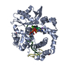
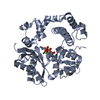
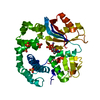
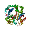
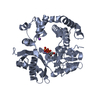
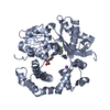
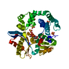
IMIDAZOLE / DNA-directed DNA/RNA polymerase mu
Similarity search - Component
Biological speciesHomo sapiens (human)
MethodX-RAY DIFFRACTION / MOLECULAR REPLACEMENT / molecular replacement / Resolution: 1.849 Å
AuthorsMoon, A.F. / Pryor, J.M. / Ramsden, D.A. / Kunkel, T.A. / Bebenek, K. / Pedersen, L.C.
CitationJournal: Nat.Struct.Mol.Biol. / Year: 2014
Title: Sustained active site rigidity during synthesis by human DNA polymerase mu.
Authors: Moon, A.F. / Pryor, J.M. / Ramsden, D.A. / Kunkel, T.A. / Bebenek, K. / Pedersen, L.C.
History
DepositionJul 31, 2013Deposition site: RCSB / Processing site: RCSB
Revision 1.0Feb 5, 2014Provider: repository / Type: Initial release
Revision 1.1Mar 5, 2014Group: Database references
Revision 1.2Mar 19, 2014Group: Database references
Revision 1.3Nov 15, 2017Group: Refinement description / Category: software
Item: _software.classification / _software.contact_author ..._software.classification / _software.contact_author / _software.contact_author_email / _software.date / _software.language / _software.location / _software.name / _software.type / _software.version
Revision 1.4Sep 20, 2023Group: Data collection / Database references ...Data collection / Database references / Derived calculations / Refinement description
Category: chem_comp_atom / chem_comp_bond ...chem_comp_atom / chem_comp_bond / database_2 / pdbx_initial_refinement_model / pdbx_struct_conn_angle / struct_conn / struct_ref_seq_dif / struct_site
Item: _database_2.pdbx_DOI / _database_2.pdbx_database_accession ..._database_2.pdbx_DOI / _database_2.pdbx_database_accession / _pdbx_struct_conn_angle.ptnr1_auth_comp_id / _pdbx_struct_conn_angle.ptnr1_auth_seq_id / _pdbx_struct_conn_angle.ptnr1_label_alt_id / _pdbx_struct_conn_angle.ptnr1_label_asym_id / _pdbx_struct_conn_angle.ptnr1_label_comp_id / _pdbx_struct_conn_angle.ptnr1_label_seq_id / _pdbx_struct_conn_angle.ptnr3_auth_comp_id / _pdbx_struct_conn_angle.ptnr3_auth_seq_id / _pdbx_struct_conn_angle.ptnr3_label_alt_id / _pdbx_struct_conn_angle.ptnr3_label_asym_id / _pdbx_struct_conn_angle.ptnr3_label_comp_id / _pdbx_struct_conn_angle.ptnr3_label_seq_id / _pdbx_struct_conn_angle.value / _struct_conn.pdbx_dist_value / _struct_conn.pdbx_ptnr2_label_alt_id / _struct_conn.ptnr1_auth_comp_id / _struct_conn.ptnr1_auth_seq_id / _struct_conn.ptnr1_label_asym_id / _struct_conn.ptnr1_label_atom_id / _struct_conn.ptnr1_label_comp_id / _struct_conn.ptnr1_label_seq_id / _struct_conn.ptnr2_auth_comp_id / _struct_conn.ptnr2_auth_seq_id / _struct_conn.ptnr2_label_asym_id / _struct_conn.ptnr2_label_atom_id / _struct_conn.ptnr2_label_comp_id / _struct_ref_seq_dif.details / _struct_site.pdbx_auth_asym_id / _struct_site.pdbx_auth_comp_id / _struct_site.pdbx_auth_seq_id

-
Structure visualization

Structure viewerMolecule:
MolmilJmol/JSmol

Downloads & links

-
Assembly

Deposited unit
A: DNA-directed DNA/RNA polymerase mu
hetero molecules


Theoretical massNumber of molelcules
Total (without water)40,2445
Polymers40,0541
Non-polymers1904
Water5,909328
1


  • Idetical with deposited unit
  • defined by author&software
TypeNameSymmetry operationNumber
identity operation1_555x,y,z1
MethodPISA
Unit cell
Length a, b, c (Å)124.192, 124.192, 50.407
Angle α, β, γ (deg.)90.000, 90.000, 90.000
Int Tables number92
Space group name H-MP41212
Components on special symmetry positions
IDModelComponents
11A-616-

HOH

21A-689-

HOH

31A-889-

HOH

-
Components

-
Protein , 1 types, 1 molecules A

#1: Protein DNA-directed DNA/RNA polymerase mu / Pol Mu / Terminal transferase


Mass: 40054.434 Da / Num. of mol.: 1
Fragment: Polymerase Mu Loop2 deletion variant, UNP residues 132-494
Source method: isolated from a genetically manipulated source
Details: Modified version of the pGEX4T3 vector backbone, where a TEV protease cleavage site has been inserted downstream of the thrombin cleavage site, and the sequence of the multicloning site has ...Details: Modified version of the pGEX4T3 vector backbone, where a TEV protease cleavage site has been inserted downstream of the thrombin cleavage site, and the sequence of the multicloning site has been replaced by that from the pMALX vector (Moon et al, 2010). Three TGA stop codons have been inserted downstream of the MCS, each utilizing a different reading frame.
Source: (gene. exp.) Homo sapiens (human) / Gene: POLM, polmu / Plasmid: pGEXM / Production host: Escherichia coli (E. coli) / References: UniProt: Q9NP87, DNA-directed DNA polymerase

-
Non-polymers , 5 types, 332 molecules

#2: Chemical ChemComp-NA / SODIUM ION


Mass: 22.990 Da / Num. of mol.: 1 / Source method: obtained synthetically / Formula: Na
#3: Chemical ChemComp-IMD / IMIDAZOLE


Mass: 69.085 Da / Num. of mol.: 1 / Source method: obtained synthetically / Formula: C3H5N2
#4: Chemical ChemComp-EDO / 1,2-ETHANEDIOL / ETHYLENE GLYCOL


Mass: 62.068 Da / Num. of mol.: 1 / Source method: obtained synthetically / Formula: C2H6O2
#5: Chemical ChemComp-CL / CHLORIDE ION


Mass: 35.453 Da / Num. of mol.: 1 / Source method: obtained synthetically / Formula: Cl
#6: Water ChemComp-HOH / water


Mass: 18.015 Da / Num. of mol.: 328 / Source method: isolated from a natural source / Formula: H2O

-
Experimental details

-
Experiment

ExperimentMethod: X-RAY DIFFRACTION / Number of used crystals: 1

-
Sample preparation

CrystalDensity Matthews: 2.43 Å3/Da / Density % sol: 49.3 %
Crystal growTemperature: 295 K / Method: vapor diffusion, hanging drop / pH: 8
Details: Crystals were grown by mixing 1 uL of concentrated protein (10.7mg/mL) with 1 uL of mother liquor (50 mM imidazole pH 8, 0.8M sodium citrate) at room temperature, using the hanging drop ...Details: Crystals were grown by mixing 1 uL of concentrated protein (10.7mg/mL) with 1 uL of mother liquor (50 mM imidazole pH 8, 0.8M sodium citrate) at room temperature, using the hanging drop vapor diffusion technique. The crystals reached usable size within 24 hours, and were then directly transferred to a cryoprotectant solution containing 50mM imidazole pH 8, 75mM NaCl, 1M sodium citrate, 10% glycerol., VAPOR DIFFUSION, HANGING DROP, temperature 295K

-
Data collection

DiffractionMean temperature: 93 K
Diffraction sourceSource: ROTATING ANODE / Type: RIGAKU MICROMAX-007 HF / Wavelength: 1.54 Å
DetectorType: RIGAKU SATURN 92 / Detector: CCD / Date: Sep 3, 2012 / Details: VariMaxHF mirrors
RadiationProtocol: SINGLE WAVELENGTH / Monochromatic (M) / Laue (L): M / Scattering type: x-ray
Radiation wavelengthWavelength: 1.54 Å / Relative weight: 1
ReflectionResolution: 1.849→50 Å / Num. all: 34121 / Num. obs: 34121 / % possible obs: 99.3 % / Observed criterion σ(F): 0 / Observed criterion σ(I): -3 / Redundancy: 9 % / Biso Wilson estimate: 22.27 Å2 / Rsym value: 0.056 / Χ2: 1.073 / Net I/σ(I): 16.4
Reflection shell
Resolution (Å)Redundancy (%)Rmerge(I) obsNum. unique allΧ2Diffraction-ID% possible all
1.85-1.924.80.41432451.047196.4
1.92-1.995.80.30833201.078198.4
1.99-2.086.10.21633421.036198.9
2.08-2.196.50.16433981.037199.9
2.19-2.337.60.13733661.0971100
2.33-2.519.10.11234211.0871100
2.51-2.76100.08834211.0911100
2.76-3.1611.80.06734581.0831100
3.16-3.9914.20.04334841.0821100
3.99-5013.50.03736661.055199.5

-
Phasing

PhasingMethod: molecular replacement

-
Processing

Software
NameVersionClassificationNB
DENZOdata reduction
SCALEPACKdata scaling
PHASERphasing
PHENIX1.8_1069refinement
PDB_EXTRACT3.11data extraction
StructureStudiodata collection
RefinementMethod to determine structure: MOLECULAR REPLACEMENT
Starting model: 2IHM (molecule B) was used as the starting molecule for molecular replacement for a lower resolution structure, which was then used as the starting model for the current higher resolution structure.
Resolution: 1.849→25.856 Å / Occupancy max: 1 / Occupancy min: 0.25 / FOM work R set: 0.8503 / SU ML: 0.19 / Cross valid method: THROUGHOUT / σ(F): 1.34 / Phase error: 21.45 / Stereochemistry target values: ML
RfactorNum. reflection% reflectionSelection details
Rfree0.2283 1708 5.01 %random
Rwork0.1809 ---
all-34121 --
obs-34070 99.34 %-
Solvent computationShrinkage radii: 0.9 Å / VDW probe radii: 1.11 Å / Solvent model: FLAT BULK SOLVENT MODEL
Displacement parametersBiso max: 69.3 Å2 / Biso mean: 19.6347 Å2 / Biso min: 7.61 Å2
Refinement stepCycle: LAST / Resolution: 1.849→25.856 Å
ProteinNucleic acidLigandSolventTotal
Num. atoms2616 0 11 328 2955
Refine LS restraints
Refine-IDTypeDev idealNumber
X-RAY DIFFRACTIONf_bond_d0.0112761
X-RAY DIFFRACTIONf_angle_d1.283759
X-RAY DIFFRACTIONf_chiral_restr0.094411
X-RAY DIFFRACTIONf_plane_restr0.006498
X-RAY DIFFRACTIONf_dihedral_angle_d14.081017
LS refinement shell

Refine-ID: X-RAY DIFFRACTION / Total num. of bins used: 12

Resolution (Å)Rfactor RfreeNum. reflection RfreeRfactor RworkNum. reflection RworkNum. reflection all% reflection obs (%)
1.8489-1.90330.30661370.2292569270696
1.9033-1.96470.30951380.21122596273497
1.9647-2.03490.25791400.19662636277699
2.0349-2.11640.25191410.178526582799100
2.1164-2.21260.24591410.17526842825100
2.2126-2.32920.23051430.1826782821100
2.3292-2.4750.23681390.194427002839100
2.475-2.6660.26231430.193527192862100
2.666-2.93390.25061440.192326912835100
2.9339-3.35770.21551450.181227522897100
3.3577-4.22730.18451440.150927672911100
4.2273-25.85890.21231530.181629123065100

+
About Yorodumi

-
News

-
Feb 9, 2022. New format data for meta-information of EMDB entries

New format data for meta-information of EMDB entries

  • Version 3 of the EMDB header file is now the official format.
  • The previous official version 1.9 will be removed from the archive.

Related info.:EMDB header

External links:wwPDB to switch to version 3 of the EMDB data model

-
Aug 12, 2020. Covid-19 info

Covid-19 info

URL: https://pdbjlvh1.pdbj.org/emnavi/covid19.php

New page: Covid-19 featured information page in EM Navigator.

Related info.:Covid-19 info / Mar 5, 2020. Novel coronavirus structure data

+
Mar 5, 2020. Novel coronavirus structure data

Novel coronavirus structure data

Related info.:Yorodumi Speices / Aug 12, 2020. Covid-19 info

External links:COVID-19 featured content - PDBj / Molecule of the Month (242):Coronavirus Proteases

+
Jan 31, 2019. EMDB accession codes are about to change! (news from PDBe EMDB page)

EMDB accession codes are about to change! (news from PDBe EMDB page)

  • The allocation of 4 digits for EMDB accession codes will soon come to an end. Whilst these codes will remain in use, new EMDB accession codes will include an additional digit and will expand incrementally as the available range of codes is exhausted. The current 4-digit format prefixed with “EMD-” (i.e. EMD-XXXX) will advance to a 5-digit format (i.e. EMD-XXXXX), and so on. It is currently estimated that the 4-digit codes will be depleted around Spring 2019, at which point the 5-digit format will come into force.
  • The EM Navigator/Yorodumi systems omit the EMD- prefix.

Related info.:Q: What is EMD? / ID/Accession-code notation in Yorodumi/EM Navigator

External links:EMDB Accession Codes are Changing Soon! / Contact to PDBj

+
Jul 12, 2017. Major update of PDB

Major update of PDB

  • wwPDB released updated PDB data conforming to the new PDBx/mmCIF dictionary.
  • This is a major update changing the version number from 4 to 5, and with Remediation, in which all the entries are updated.
  • In this update, many items about electron microscopy experimental information are reorganized (e.g. em_software).
  • Now, EM Navigator and Yorodumi are based on the updated data.

External links:wwPDB Remediation / Enriched Model Files Conforming to OneDep Data Standards Now Available in the PDB FTP Archive

-
Yorodumi

Thousand views of thousand structures

  • Yorodumi is a browser for structure data from EMDB, PDB, SASBDB, etc.
  • This page is also the successor to EM Navigator detail page, and also detail information page/front-end page for Omokage search.
  • The word "yorodu" (or yorozu) is an old Japanese word meaning "ten thousand". "mi" (miru) is to see.

Related info.:EMDB / PDB / SASBDB / Comparison of 3 databanks / Yorodumi Search / Aug 31, 2016. New EM Navigator & Yorodumi / Yorodumi Papers / Jmol/JSmol / Function and homology information / Changes in new EM Navigator and Yorodumi

Read more