- PDB-5v70: Crystal structure of N102A mutant of human macrophage migration i... -
+
Open data
ID or keywords:
Loading...
-
Basic information
Entry
Database: PDB / ID: 5v70
Title
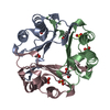
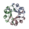
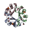
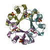
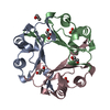
Crystal structure of N102A mutant of human macrophage migration inhibitory factor
Components
Macrophage migration inhibitory factor
Keywords
ISOMERASE / trimeric / mutation
Function / homology
Function and homology information
: / positive regulation of myeloid leukocyte cytokine production involved in immune response / phenylpyruvate tautomerase / L-dopachrome isomerase / regulation of macrophage activation / dopachrome isomerase activity / phenylpyruvate tautomerase activity / cytokine receptor binding / negative regulation of myeloid cell apoptotic process / negative regulation of mature B cell apoptotic process ...: / positive regulation of myeloid leukocyte cytokine production involved in immune response / phenylpyruvate tautomerase / L-dopachrome isomerase / regulation of macrophage activation / dopachrome isomerase activity / phenylpyruvate tautomerase activity / cytokine receptor binding / negative regulation of myeloid cell apoptotic process / negative regulation of mature B cell apoptotic process / negative regulation of macrophage chemotaxis / carboxylic acid metabolic process / positive regulation of arachidonate secretion / positive regulation of lipopolysaccharide-mediated signaling pathway / prostaglandin biosynthetic process / positive regulation of chemokine (C-X-C motif) ligand 2 production / negative regulation of protein metabolic process / negative regulation of intrinsic apoptotic signaling pathway in response to DNA damage by p53 class mediator / protein homotrimerization / chemoattractant activity / negative regulation of DNA damage response, signal transduction by p53 class mediator / negative regulation of cellular senescence / positive regulation of cAMP/PKA signal transduction / positive regulation of phosphorylation / positive regulation of B cell proliferation / Gene and protein expression by JAK-STAT signaling after Interleukin-12 stimulation / negative regulation of cell migration / cytokine activity / positive regulation of cytokine production / Cell surface interactions at the vascular wall / DNA damage response, signal transduction by p53 class mediator / positive regulation of fibroblast proliferation / cellular senescence / positive regulation of tumor necrosis factor production / protease binding / secretory granule lumen / vesicle / ficolin-1-rich granule lumen / positive regulation of ERK1 and ERK2 cascade / cell surface receptor signaling pathway / inflammatory response / negative regulation of gene expression / innate immune response / positive regulation of cell population proliferation / Neutrophil degranulation / negative regulation of apoptotic process / cell surface / : / extracellular exosome / extracellular region / nucleoplasm / identical protein binding / plasma membrane / cytosol / cytoplasm Similarity search - Function
Component-ID: _ / Beg auth comp-ID: PRO / Beg label comp-ID: PRO / End auth comp-ID: ALA / End label comp-ID: ALA / Refine code: _ / Auth seq-ID: 1 - 114 / Label seq-ID: 1 - 114
 Movie
Movie Controller

 Yorodumi
 Open data
 Basic information
 Components
 Keywords
 Function and homology information
 Homo sapiens (human)
 X-RAY DIFFRACTION /
  Authors
 Citation
 Structure visualization
 Downloads & links
 Other downloads


 PDBj
PDBj


 Assembly
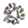


 Mass: 96.063 Da / Num. of mol.: 5 / Source method: obtained synthetically / Formula: SO4
 Mass: 92.094 Da / Num. of mol.: 3 / Source method: obtained synthetically / Formula: C3H8O3
 Mass: 60.095 Da / Num. of mol.: 3 / Source method: obtained synthetically / Formula: C3H8O
 Mass: 18.015 Da / Num. of mol.: 214 / Source method: isolated from a natural source / Formula: H2O
 Sample preparation
 Processing