 Movie
Movie Controller

[English] 日本語
 Yorodumi
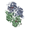
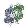
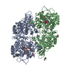
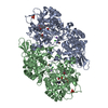
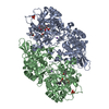
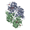
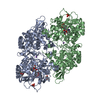
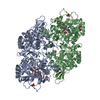
- PDB-5v53: Crystal structure of the D141A/Q233E/N240D variant of catalase-pe... -
+ Open data

- Basic information
| Entry | Database: PDB / ID: 5v53 | ||||||
|---|---|---|---|---|---|---|---|
| Title | Crystal structure of the D141A/Q233E/N240D variant of catalase-peroxidase from B. pseudomallei with acetate bound | ||||||
 Components Components | Catalase-peroxidase | ||||||
 Keywords Keywords | OXIDOREDUCTASE / catalase / peroxidase / D141A/Q233E/N240D / heme / acetate | ||||||
| Function / homology |  Function and homology information Function and homology informationcatalase-peroxidase / catalase activity / hydrogen peroxide catabolic process / cellular response to hydrogen peroxide / heme binding / metal ion binding / cytosol Similarity search - Function | ||||||
| Biological species |  Burkholderia pseudomallei (bacteria) Burkholderia pseudomallei (bacteria) | ||||||
| Method |  X-RAY DIFFRACTION / SYNCHROTRON / MOLECULAR REPLACEMENT / Resolution: 1.7 Å X-RAY DIFFRACTION / SYNCHROTRON / MOLECULAR REPLACEMENT / Resolution: 1.7 Å | ||||||
 Authors Authors | Loewen, P.C. | ||||||
| Funding support |  Canada, 1items Canada, 1items
| ||||||
 Citation Citation | Journal: Biochemistry / Year: 2017 Title: The Catalase Activity of Catalase-Peroxidases Is Modulated by Changes in the pKa of the Distal Histidine. Authors: Machuqueiro, M. / Victor, B. / Switala, J. / Villanueva, J. / Rovira, C. / Fita, I. / Loewen, P.C. | ||||||
| History |
|
- Structure visualization
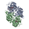
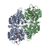
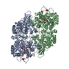
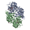
| Structure viewer | Molecule: MolmilJmol/JSmol |
|---|
- Downloads & links
-Download
| PDBx/mmCIF format | 5v53.cif.gz | 575.6 KB | Display | PDBx/mmCIF format |
|---|---|---|---|---|
| PDB format | pdb5v53.ent.gz | 466.3 KB | Display | PDB format |
| PDBx/mmJSON format | 5v53.json.gz | Tree view | PDBx/mmJSON format | |
| Others |  Other downloads Other downloads |
-Validation report
| Arichive directory | https://data.pdbj.org/pub/pdb/validation_reports/v5/5v53ftp://data.pdbj.org/pub/pdb/validation_reports/v5/5v53 | HTTPS FTP |
|---|
-Related structure data
| Related structure data |  5kq0C  5kq2C  5kq3C  5kq6C  5kqhC  5kqiC  5kqkC  5ksfC  5kskC  5syuC  5syvC  5sywC  5txqC  5v4oC  1mwv C: citing same article ( S: Starting model for refinement |
|---|---|
| Similar structure data |


-Links
 PDBj
PDBj


- Assembly
| Deposited unit | 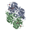
| ||||||||||||||||||
|---|---|---|---|---|---|---|---|---|---|---|---|---|---|---|---|---|---|---|---|
| 1 |
| ||||||||||||||||||
| Unit cell |
| ||||||||||||||||||
| Noncrystallographic symmetry (NCS) | NCS domain:
NCS domain segments: Component-ID: _ / Ens-ID: 1 / Beg auth comp-ID: GLY / Beg label comp-ID: GLY / End auth comp-ID: ALA / End label comp-ID: ALA / Refine code: _ / Auth seq-ID: 36 - 748 / Label seq-ID: 16 - 728
|
-Components
-Protein , 1 types, 2 molecules AB
| #1: Protein | Mass: 79494.945 Da / Num. of mol.: 2 / Mutation: D141A, Q233E, N240D Source method: isolated from a genetically manipulated source Source: (gene. exp.) Burkholderia pseudomallei (strain 1710b) (bacteria)Strain: 1710b / Gene: katG, BURPS1710b_3366 / Plasmid: pKS / Production host: |
|---|
-Non-polymers , 8 types, 1481 molecules 


| #2: Chemical |  Mass: 616.487 Da / Num. of mol.: 2 / Source method: obtained synthetically / Formula: C34H32FeN4O4 Mass: 616.487 Da / Num. of mol.: 2 / Source method: obtained synthetically / Formula: C34H32FeN4O4#3: Chemical |  Mass: 22.990 Da / Num. of mol.: 2 / Source method: obtained synthetically / Formula: Na Mass: 22.990 Da / Num. of mol.: 2 / Source method: obtained synthetically / Formula: Na#4: Chemical |  Mass: 35.453 Da / Num. of mol.: 2 / Source method: obtained synthetically / Formula: Cl Mass: 35.453 Da / Num. of mol.: 2 / Source method: obtained synthetically / Formula: Cl#5: Chemical |  Mass: 31.999 Da / Num. of mol.: 2 / Source method: obtained synthetically / Formula: O2 Mass: 31.999 Da / Num. of mol.: 2 / Source method: obtained synthetically / Formula: O2#6: Chemical |  Mass: 59.044 Da / Num. of mol.: 2 / Source method: obtained synthetically / Formula: C2H3O2 Mass: 59.044 Da / Num. of mol.: 2 / Source method: obtained synthetically / Formula: C2H3O2#7: Chemical |  Mass: 94.971 Da / Num. of mol.: 2 / Source method: obtained synthetically / Formula: PO4 Mass: 94.971 Da / Num. of mol.: 2 / Source method: obtained synthetically / Formula: PO4#8: Chemical | ChemComp-MPD / (  Mass: 118.174 Da / Num. of mol.: 4 / Source method: obtained synthetically / Formula: C6H14O2 / Comment: precipitant*YM Mass: 118.174 Da / Num. of mol.: 4 / Source method: obtained synthetically / Formula: C6H14O2 / Comment: precipitant*YM#9: Water | ChemComp-HOH / | Mass: 18.015 Da / Num. of mol.: 1465 / Source method: isolated from a natural source / Formula: H2O |
|---|
-Details
| Has protein modification | Y |
|---|
-Experimental details
-Experiment
| Experiment | Method: X-RAY DIFFRACTION / Number of used crystals: 1 |
|---|
- Sample preparation
| Crystal | Density Matthews: 3.28 Å3/Da / Density % sol: 62.45 % / Mosaicity: 0.17 ° |
|---|---|
| Crystal grow | Temperature: 293 K / Method: vapor diffusion, hanging drop / pH: 6.1 / Details: MPD |
-Data collection
| Diffraction | Mean temperature: 100 K | |||||||||||||||||||||||||||||||||||||||||||||||||||||||||||||||||||||||||||||||||||||||||||||||||||
|---|---|---|---|---|---|---|---|---|---|---|---|---|---|---|---|---|---|---|---|---|---|---|---|---|---|---|---|---|---|---|---|---|---|---|---|---|---|---|---|---|---|---|---|---|---|---|---|---|---|---|---|---|---|---|---|---|---|---|---|---|---|---|---|---|---|---|---|---|---|---|---|---|---|---|---|---|---|---|---|---|---|---|---|---|---|---|---|---|---|---|---|---|---|---|---|---|---|---|---|---|
| Diffraction source | Source: SYNCHROTRON / Site: CLSI / Beamline: 08ID-1 / Wavelength: 0.98 Å | |||||||||||||||||||||||||||||||||||||||||||||||||||||||||||||||||||||||||||||||||||||||||||||||||||
| Detector | Type: MARMOSAIC 300 mm CCD / Detector: CCD / Date: Oct 9, 2015 / Details: mirrors | |||||||||||||||||||||||||||||||||||||||||||||||||||||||||||||||||||||||||||||||||||||||||||||||||||
| Radiation | Monochromator: DOUBLE CRYSTAL / Protocol: SINGLE WAVELENGTH / Monochromatic (M) / Laue (L): M / Scattering type: x-ray | |||||||||||||||||||||||||||||||||||||||||||||||||||||||||||||||||||||||||||||||||||||||||||||||||||
| Radiation wavelength | Wavelength: 0.98 Å / Relative weight: 1 | |||||||||||||||||||||||||||||||||||||||||||||||||||||||||||||||||||||||||||||||||||||||||||||||||||
| Reflection | Resolution: 1.7→46.23 Å / Num. all: 214623 / Num. obs: 214623 / % possible obs: 96.1 % / Redundancy: 3.9 % / Rpim(I) all: 0.034 / Rrim(I) all: 0.07 / Rsym value: 0.061 / Net I/av σ(I): 8.6 / Net I/σ(I): 11.7 / Num. measured all: 827169 | |||||||||||||||||||||||||||||||||||||||||||||||||||||||||||||||||||||||||||||||||||||||||||||||||||
| Reflection shell | Diffraction-ID: 1
|
- Processing
| Software |
| |||||||||||||||||||||||||||||||||||||||||||||||||||||||||||||||||||||||||||
|---|---|---|---|---|---|---|---|---|---|---|---|---|---|---|---|---|---|---|---|---|---|---|---|---|---|---|---|---|---|---|---|---|---|---|---|---|---|---|---|---|---|---|---|---|---|---|---|---|---|---|---|---|---|---|---|---|---|---|---|---|---|---|---|---|---|---|---|---|---|---|---|---|---|---|---|---|
| Refinement | Method to determine structure: MOLECULAR REPLACEMENT Starting model: 1MWV 1mwv Resolution: 1.7→46.23 Å / Cor.coef. Fo:Fc: 0.977 / Cor.coef. Fo:Fc free: 0.968 / SU B: 3.09 / SU ML: 0.052 / SU R Cruickshank DPI: 0.0718 / Cross valid method: THROUGHOUT / σ(F): 0 / ESU R: 0.072 / ESU R Free: 0.075 / Details: MOLECULAR REPLACEMENT
| |||||||||||||||||||||||||||||||||||||||||||||||||||||||||||||||||||||||||||
| Solvent computation | Ion probe radii: 0.8 Å / Shrinkage radii: 0.8 Å / VDW probe radii: 1.2 Å | |||||||||||||||||||||||||||||||||||||||||||||||||||||||||||||||||||||||||||
| Displacement parameters | Biso max: 111.9 Å2 / Biso mean: 25.554 Å2 / Biso min: 11.63 Å2
| |||||||||||||||||||||||||||||||||||||||||||||||||||||||||||||||||||||||||||
| Refinement step | Cycle: final / Resolution: 1.7→46.23 Å
| |||||||||||||||||||||||||||||||||||||||||||||||||||||||||||||||||||||||||||
| Refine LS restraints |
| |||||||||||||||||||||||||||||||||||||||||||||||||||||||||||||||||||||||||||
| Refine LS restraints NCS | Ens-ID: 1 / Number: 47218 / Refine-ID: X-RAY DIFFRACTION / Type: interatomic distance / Rms dev position: 0.07 Å / Weight position: 0.05
| |||||||||||||||||||||||||||||||||||||||||||||||||||||||||||||||||||||||||||
| LS refinement shell | Resolution: 1.7→1.744 Å / Rfactor Rfree error: 0 / Total num. of bins used: 20
| |||||||||||||||||||||||||||||||||||||||||||||||||||||||||||||||||||||||||||
| Refinement TLS params. | Method: refined / Refine-ID: X-RAY DIFFRACTION
| |||||||||||||||||||||||||||||||||||||||||||||||||||||||||||||||||||||||||||
| Refinement TLS group |
|
