| Entry | Database: PDB / ID: 5v2w
|
|---|
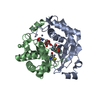
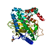
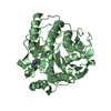
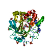
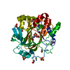
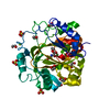
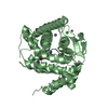
| Title | Crystal structure of a LuxS from salmonella typhi |
|---|
 Components Components | S-ribosylhomocysteine lyase |
|---|
 Keywords Keywords | LYASE / Quroum sensing protein / LuxS / Salmonella typhi / S-ribosylhomocysteine lyase |
|---|
| Function / homology |  Function and homology information Function and homology information
S-ribosylhomocysteinase (LuxS) / S-ribosylhomocysteinase (LuxS) / S-ribosylhomocysteinase (LuxS) superfamily / S-Ribosylhomocysteinase (LuxS) / Metalloenzyme, LuxS/M16 peptidase-like / Gyrase A; domain 2 / 2-Layer Sandwich / Alpha BetaSimilarity search - Domain/homology |
|---|
| Biological species |  Salmonella typhi (bacteria) Salmonella typhi (bacteria) |
|---|
| Method |  X-RAY DIFFRACTION / MOLECULAR REPLACEMENT / Resolution: 2.3 Å X-RAY DIFFRACTION / MOLECULAR REPLACEMENT / Resolution: 2.3 Å |
|---|
 Authors Authors | Perumal, P. / Raina, R. / Manoj Kumar, P. / Arockisamy, A. / SundaraBaalaji, N. |
|---|
| Funding support |  India, 1items India, 1items | Organization | Grant number | Country |
|---|
| ICMR | 2011-15260 | India |
|
|---|
 Citation Citation | Journal: To Be Published
Title: Crystal structure of a LuxS from salmonella typhi
Authors: Perumal, P. / Raina, R. / Arockisamy, A. / SundaraBaalaji, N. |
|---|
| History | | Deposition | Mar 6, 2017 | Deposition site: RCSB / Processing site: PDBJ |
|---|
| Revision 1.0 | Aug 23, 2017 | Provider: repository / Type: Initial release |
|---|
| Revision 1.1 | Sep 27, 2017 | Group: Data collection / Category: diffrn_detector / Item: _diffrn_detector.detector |
|---|
| Revision 1.2 | Nov 8, 2023 | Group: Data collection / Database references ...Data collection / Database references / Derived calculations / Refinement description
Category: chem_comp_atom / chem_comp_bond ...chem_comp_atom / chem_comp_bond / database_2 / pdbx_initial_refinement_model / struct_conn / struct_conn_type / struct_ncs_dom_lim
Item: _database_2.pdbx_DOI / _database_2.pdbx_database_accession ..._database_2.pdbx_DOI / _database_2.pdbx_database_accession / _struct_conn.conn_type_id / _struct_conn.id / _struct_conn.pdbx_dist_value / _struct_conn.pdbx_leaving_atom_flag / _struct_conn.ptnr1_auth_asym_id / _struct_conn.ptnr1_auth_comp_id / _struct_conn.ptnr1_auth_seq_id / _struct_conn.ptnr1_label_asym_id / _struct_conn.ptnr1_label_atom_id / _struct_conn.ptnr1_label_comp_id / _struct_conn.ptnr1_label_seq_id / _struct_conn.ptnr2_auth_asym_id / _struct_conn.ptnr2_auth_comp_id / _struct_conn.ptnr2_auth_seq_id / _struct_conn.ptnr2_label_asym_id / _struct_conn.ptnr2_label_atom_id / _struct_conn.ptnr2_label_comp_id / _struct_conn.ptnr2_label_seq_id / _struct_conn_type.id / _struct_ncs_dom_lim.beg_auth_comp_id / _struct_ncs_dom_lim.beg_label_asym_id / _struct_ncs_dom_lim.beg_label_comp_id / _struct_ncs_dom_lim.beg_label_seq_id / _struct_ncs_dom_lim.end_auth_comp_id / _struct_ncs_dom_lim.end_label_asym_id / _struct_ncs_dom_lim.end_label_comp_id / _struct_ncs_dom_lim.end_label_seq_id |
|---|
| Revision 1.3 | Oct 30, 2024 | Group: Structure summary / Category: pdbx_entry_details / pdbx_modification_feature |
|---|
|
|---|
 Movie
Movie Controller


 Open data
 Basic information
 Structure visualization
 Downloads & links
 Other downloads


 PDBj
PDBj

 Assembly
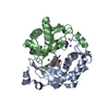

 Mass: 65.409 Da / Num. of mol.: 2 / Source method: isolated from a natural source / Formula: Zn
 Mass: 18.015 Da / Num. of mol.: 42 / Source method: isolated from a natural source / Formula: H2O
 Sample preparation
 Processing