 Movie
Movie Controller

[English] 日本語
 Yorodumi
- PDB-5uun: Crystal structure of SARO_2595 from Novosphingobium aromaticivorans -
+ Open data

- Basic information
| Entry | Database: PDB / ID: 5uun | ||||||
|---|---|---|---|---|---|---|---|
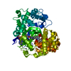
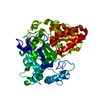
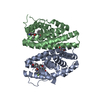
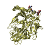
| Title | Crystal structure of SARO_2595 from Novosphingobium aromaticivorans | ||||||
 Components Components | Glutathione S-transferase-like protein | ||||||
 Keywords Keywords | TRANSFERASE / Bioenergy / GLBRC / lignin valorization | ||||||
| Function / homology |  Function and homology information Function and homology information | ||||||
| Biological species |  Novosphingobium aromaticivorans (bacteria) Novosphingobium aromaticivorans (bacteria) | ||||||
| Method |  X-RAY DIFFRACTION / SYNCHROTRON / MOLECULAR REPLACEMENT / Resolution: 1.45 Å X-RAY DIFFRACTION / SYNCHROTRON / MOLECULAR REPLACEMENT / Resolution: 1.45 Å | ||||||
 Authors Authors | Bingman, C.A. / Kontur, W.S. / Olmsted, C.N. / Fox, B.G. / Donohue, T.J. | ||||||
 Citation Citation | Journal: J. Biol. Chem. / Year: 2018 Title: Novosphingobium aromaticivoransuses a Nu-class glutathioneS-transferase as a glutathione lyase in breaking the beta-aryl ether bond of lignin. Authors: Kontur, W.S. / Bingman, C.A. / Olmsted, C.N. / Wassarman, D.R. / Ulbrich, A. / Gall, D.L. / Smith, R.W. / Yusko, L.M. / Fox, B.G. / Noguera, D.R. / Coon, J.J. / Donohue, T.J. | ||||||
| History |
|
- Structure visualization
| Structure viewer | Molecule: MolmilJmol/JSmol |
|---|
- Downloads & links
-Download
| PDBx/mmCIF format | 5uun.cif.gz | 373.6 KB | Display | PDBx/mmCIF format |
|---|---|---|---|---|
| PDB format | pdb5uun.ent.gz | 313.7 KB | Display | PDB format |
| PDBx/mmJSON format | 5uun.json.gz | Tree view | PDBx/mmJSON format | |
| Others |  Other downloads Other downloads |
-Validation report
| Arichive directory | https://data.pdbj.org/pub/pdb/validation_reports/uu/5uunftp://data.pdbj.org/pub/pdb/validation_reports/uu/5uun | HTTPS FTP |
|---|
-Related structure data
| Related structure data |  5uuoC  3c8eS C: citing same article ( S: Starting model for refinement |
|---|---|
| Similar structure data |


-Links
 PDBj
PDBj

- Assembly
| Deposited unit | 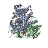
| ||||||||
|---|---|---|---|---|---|---|---|---|---|
| 1 |
| ||||||||
| Unit cell |
|
-Components
| #1: Protein | Mass: 32515.617 Da / Num. of mol.: 2 Source method: isolated from a genetically manipulated source Source: (gene. exp.) Novosphingobium aromaticivorans (strain ATCC 700278 / DSM 12444 / CIP 105152 / NBRC 16084 / F199) (bacteria)Strain: ATCC 700278 / DSM 12444 / CIP 105152 / NBRC 16084 / F199 Gene: Saro_2595 / Cell line (production host): B834 / Production host: #2: Chemical | ChemComp-GSH /   Mass: 307.323 Da / Num. of mol.: 4 / Source method: obtained synthetically / Formula: C10H17N3O6S Mass: 307.323 Da / Num. of mol.: 4 / Source method: obtained synthetically / Formula: C10H17N3O6S#3: Chemical | ChemComp-ACT /   Mass: 59.044 Da / Num. of mol.: 21 / Source method: obtained synthetically / Formula: C2H3O2 Mass: 59.044 Da / Num. of mol.: 21 / Source method: obtained synthetically / Formula: C2H3O2#4: Water | ChemComp-HOH / |  Mass: 18.015 Da / Num. of mol.: 972 / Source method: isolated from a natural source / Formula: H2O Mass: 18.015 Da / Num. of mol.: 972 / Source method: isolated from a natural source / Formula: H2O |
|---|
-Experimental details
-Experiment
| Experiment | Method: X-RAY DIFFRACTION / Number of used crystals: 1 |
|---|
- Sample preparation
| Crystal | Density Matthews: 3.1 Å3/Da / Density % sol: 60.38 % |
|---|---|
| Crystal grow | Temperature: 293 K / Method: vapor diffusion, sitting drop Details: Saro protein at 277 mM was incubated with 10 mM reduced glutathione for 50 minutes prior to setting up crystallization experiments. An equal volume of protein solution and reservoir were ...Details: Saro protein at 277 mM was incubated with 10 mM reduced glutathione for 50 minutes prior to setting up crystallization experiments. An equal volume of protein solution and reservoir were deposited by a TTP Labtech Mosquito in a SD2 crystallization plate and allowed to equilibrate. The crystals were self-cryoprotected by the reservoir of 4 M ammonium acetate. |
-Data collection
| Diffraction | Mean temperature: 100 K |
|---|---|
| Diffraction source | Source: SYNCHROTRON / Site: APS  / Beamline: 23-ID-B / Wavelength: 1.033 Å / Beamline: 23-ID-B / Wavelength: 1.033 Å |
| Detector | Type: DECTRIS EIGER X 16M / Detector: PIXEL / Date: Jun 30, 2016 |
| Radiation | Monochromator: Silicon 111 / Protocol: SINGLE WAVELENGTH / Monochromatic (M) / Laue (L): M / Scattering type: x-ray |
| Radiation wavelength | Wavelength: 1.033 Å / Relative weight: 1 |
| Reflection | Resolution: 1.45→43.97 Å / Num. obs: 140421 / % possible obs: 96.48 % / Redundancy: 13.2 % / Biso Wilson estimate: 13.23 Å2 / CC1/2: 1 / Rmerge(I) obs: 0.04367 / Rpim(I) all: 0.01239 / Net I/σ(I): 36.36 |
| Reflection shell | Resolution: 1.45→1.502 Å / Redundancy: 10.3 % / Rmerge(I) obs: 0.2047 / Mean I/σ(I) obs: 8.82 / Num. unique obs: 11067 / CC1/2: 0.986 / Rpim(I) all: 0.06599 / % possible all: 76.88 |
- Processing
| Software |
| ||||||||||||||||||||||||||||||||||||||||||||||||||||||||||||||||||||||||||||||||||||||||||||||||||
|---|---|---|---|---|---|---|---|---|---|---|---|---|---|---|---|---|---|---|---|---|---|---|---|---|---|---|---|---|---|---|---|---|---|---|---|---|---|---|---|---|---|---|---|---|---|---|---|---|---|---|---|---|---|---|---|---|---|---|---|---|---|---|---|---|---|---|---|---|---|---|---|---|---|---|---|---|---|---|---|---|---|---|---|---|---|---|---|---|---|---|---|---|---|---|---|---|---|---|---|
| Refinement | Method to determine structure: MOLECULAR REPLACEMENT Starting model: 3c8e chain A Resolution: 1.45→43.975 Å / SU ML: 0.08 / Cross valid method: FREE R-VALUE / σ(F): 1.35 / Phase error: 11.39 / Stereochemistry target values: ML
| ||||||||||||||||||||||||||||||||||||||||||||||||||||||||||||||||||||||||||||||||||||||||||||||||||
| Solvent computation | Shrinkage radii: 0.9 Å / VDW probe radii: 1.11 Å / Solvent model: FLAT BULK SOLVENT MODEL | ||||||||||||||||||||||||||||||||||||||||||||||||||||||||||||||||||||||||||||||||||||||||||||||||||
| Refinement step | Cycle: LAST / Resolution: 1.45→43.975 Å
| ||||||||||||||||||||||||||||||||||||||||||||||||||||||||||||||||||||||||||||||||||||||||||||||||||
| Refine LS restraints |
| ||||||||||||||||||||||||||||||||||||||||||||||||||||||||||||||||||||||||||||||||||||||||||||||||||
| LS refinement shell |
|
