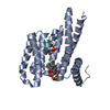
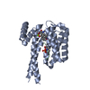
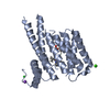
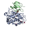
Entry Database : PDB / ID : 5uosTitle Crystal Structure of CblC (MMACHC) (1-238), a human B12 processing enzyme, complexed with an Antivitamin B12 Methylmalonic aciduria and homocystinuria type C protein Keywords / / / / Function / homology Function Domain/homology Component
/ / / / / / / / / / / / / / / / / / / / / / / / / / / / / / Biological species Homo sapiens (human)Method / / / Resolution : 2.51 Å Authors Shanmuganathan, A. / Karasik, A. / Ruetz, M. / Banerjee, R. / Krautler, B. / Koutmos, M. Funding support Organization Grant number Country American Heart Association 13SDG14560056
Journal : Angew. Chem. Int. Ed. Engl. / Year : 2017Title : Antivitamin B12 Inhibition of the Human B12 -Processing Enzyme CblC: Crystal Structure of an Inactive Ternary Complex with Glutathione as the Cosubstrate.Authors : Ruetz, M. / Shanmuganathan, A. / Gherasim, C. / Karasik, A. / Salchner, R. / Kieninger, C. / Wurst, K. / Banerjee, R. / Koutmos, M. / Krautler, B. History Deposition Feb 1, 2017 Deposition site / Processing site Revision 1.0 Jun 7, 2017 Provider / Type Revision 1.1 Jun 21, 2017 Group / Category Item _citation.country / _citation.journal_id_ASTM ... _citation.country / _citation.journal_id_ASTM / _citation.journal_id_CSD / _citation.journal_volume / _citation.page_first / _citation.page_last Revision 1.2 Sep 27, 2017 Group / Category / Item Revision 1.3 Nov 1, 2017 Group / Category Revision 1.4 Oct 4, 2023 Group / Database references / Refinement descriptionCategory chem_comp_atom / chem_comp_bond ... chem_comp_atom / chem_comp_bond / database_2 / pdbx_initial_refinement_model Item / _database_2.pdbx_database_accession
Show all Show less
 Movie
Movie Controller

 Yorodumi
 Open data
 Basic information
 Components
 Keywords
 Function and homology information
 Homo sapiens (human)
 X-RAY DIFFRACTION /
  Authors
 United States, 1items
  Citation
 Structure visualization
 Downloads & links
 Other downloads
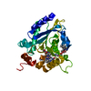

 PDBj
PDBj

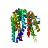
 Assembly
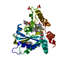


 Mass: 1330.356 Da / Num. of mol.: 1 / Source method: obtained synthetically / Formula: C62H89CoN13O14P
 Mass: 138.114 Da / Num. of mol.: 1 / Source method: obtained synthetically / Formula: C8H4F2
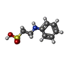
 Mass: 307.323 Da / Num. of mol.: 1 / Source method: obtained synthetically / Formula: C10H17N3O6S
 Mass: 201.243 Da / Num. of mol.: 1 / Source method: obtained synthetically / Formula: C8H11NO3S
 Mass: 96.063 Da / Num. of mol.: 2 / Source method: obtained synthetically / Formula: SO4
 Mass: 22.990 Da / Num. of mol.: 1 / Source method: obtained synthetically / Formula: Na
 Sample preparation
 Processing