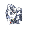
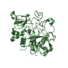
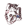
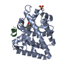
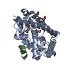
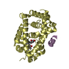
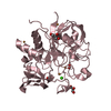
[English] 日本語
Yorodumi
- PDB-5unj: Structure of Human Liver Receptor Homolog 1 in complex with PGC1a... -

+
Open data


ID or keywords:

Loading...

-
Basic information

Entry
Database: PDB / ID: 5unj
TitleStructure of Human Liver Receptor Homolog 1 in complex with PGC1a and RJW100
Components
  • Nuclear receptor subfamily 5 group A member 2
  • Peroxisome proliferator-activated gamma coactivator 1-alpha
KeywordsNUCLEAR PROTEIN / Nuclear receptor / agonist / coregulator
Function / homology
Function and homology information


Regulation of gene expression in early pancreatic precursor cells / positive regulation of mitochondrial DNA metabolic process / positive regulation of muscle tissue development / pancreas morphogenesis / calcineurin-mediated signaling / positive regulation of cellular respiration / acinar cell differentiation / positive regulation of fatty acid oxidation / tissue development / : ...Regulation of gene expression in early pancreatic precursor cells / positive regulation of mitochondrial DNA metabolic process / positive regulation of muscle tissue development / pancreas morphogenesis / calcineurin-mediated signaling / positive regulation of cellular respiration / acinar cell differentiation / positive regulation of fatty acid oxidation / tissue development / : / bile acid metabolic process / : / lncRNA binding / cellular respiration / response to muscle activity / embryo development ending in birth or egg hatching / Activation of PPARGC1A (PGC-1alpha) by phosphorylation / temperature homeostasis / homeostatic process / positive regulation of ATP biosynthetic process / response to starvation / intracellular glucose homeostasis / fatty acid oxidation / response to dietary excess / adipose tissue development / positive regulation of viral genome replication / FOXO-mediated transcription of oxidative stress, metabolic and neuronal genes / energy homeostasis / brown fat cell differentiation / positive regulation of gluconeogenesis / digestion / respiratory electron transport chain / cellular response to leukemia inhibitory factor / hormone-mediated signaling pathway / mitochondrion organization / SUMOylation of transcription cofactors / nuclear receptor coactivator activity / RNA splicing / cholesterol homeostasis / gluconeogenesis / transcription coregulator binding / transcription initiation at RNA polymerase II promoter / nuclear receptor binding / transcription coregulator activity / negative regulation of smooth muscle cell proliferation / phospholipid binding / circadian regulation of gene expression / Heme signaling / SUMOylation of intracellular receptors / Transcriptional activation of mitochondrial biogenesis / regulation of circadian rhythm / PPARA activates gene expression / PML body / chromatin DNA binding / mRNA processing / Transcriptional regulation of white adipocyte differentiation / Nuclear Receptor transcription pathway / RNA polymerase II transcription regulator complex / Regulation of RUNX2 expression and activity / nuclear receptor activity / positive regulation of DNA-binding transcription factor activity / Circadian Clock / sequence-specific double-stranded DNA binding / cellular response to oxidative stress / positive regulation of cold-induced thermogenesis / regulation of cell population proliferation / DNA-binding transcription activator activity, RNA polymerase II-specific / protein-containing complex assembly / neuron apoptotic process / DNA-binding transcription factor binding / Estrogen-dependent gene expression / negative regulation of neuron apoptotic process / RNA polymerase II-specific DNA-binding transcription factor binding / sequence-specific DNA binding / transcription coactivator activity / protein stabilization / transcription cis-regulatory region binding / DNA-binding transcription factor activity, RNA polymerase II-specific / RNA polymerase II cis-regulatory region sequence-specific DNA binding / DNA-binding transcription factor activity / ubiquitin protein ligase binding / chromatin binding / chromatin / positive regulation of gene expression / regulation of DNA-templated transcription / regulation of transcription by RNA polymerase II / positive regulation of DNA-templated transcription / positive regulation of transcription by RNA polymerase II / DNA binding / RNA binding / zinc ion binding / nucleoplasm / nucleus / cytosol / cytoplasm
Similarity search - Function
PGC-1alpha, RNA recognition motif / PGC-1 / Nuclear hormone receptor family 5 / Retinoid X Receptor / Retinoid X Receptor / RNA recognition motif / RNA recognition motif / Eukaryotic RNA Recognition Motif (RRM) profile. / RNA recognition motif domain / RNA-binding domain superfamily ...PGC-1alpha, RNA recognition motif / PGC-1 / Nuclear hormone receptor family 5 / Retinoid X Receptor / Retinoid X Receptor / RNA recognition motif / RNA recognition motif / Eukaryotic RNA Recognition Motif (RRM) profile. / RNA recognition motif domain / RNA-binding domain superfamily / Nuclear hormone receptor / Nuclear hormones receptors DNA-binding region signature. / Zinc finger, nuclear hormone receptor-type / Zinc finger, C4 type (two domains) / Nuclear hormone receptors DNA-binding domain profile. / c4 zinc finger in nuclear hormone receptors / Nuclear hormone receptor, ligand-binding domain / Nuclear hormone receptor-like domain superfamily / Ligand-binding domain of nuclear hormone receptor / Nuclear receptor (NR) ligand-binding (LBD) domain profile. / Ligand binding domain of hormone receptors / Zinc finger, NHR/GATA-type / Nucleotide-binding alpha-beta plait domain superfamily / Orthogonal Bundle / Mainly Alpha
Similarity search - Domain/homology
Chem-RJW / Nuclear receptor subfamily 5 group A member 2 / Peroxisome proliferator-activated receptor gamma coactivator 1-alpha
Similarity search - Component
Biological speciesHomo sapiens (human)
MethodX-RAY DIFFRACTION / SYNCHROTRON / MOLECULAR REPLACEMENT / Resolution: 1.959 Å
AuthorsMays, S.G. / Ortlund, E.A.
Funding support United States, 4items
OrganizationGrant numberCountry
National Institutes of Health/National Institute of Diabetes and Digestive and Kidney Disease (NIH/NIDDK)R01DK095750 United States
National Institutes of Health/National Institute of Diabetes and Digestive and Kidney Disease (NIH/NIDDK)F31DK111171 United States
National Institutes of Health/National Institute of General Medical Sciences (NIH/NIGMS)T32GM008602 United States
National Institutes of Health/National Institute of General Medical Sciences (NIH/NIGMS)NIH K12GM000680 United States
CitationJournal: Mol. Pharmacol. / Year: 2017
Title: Structure and Dynamics of the Liver Receptor Homolog 1-PGC1 alpha Complex.
Authors: Mays, S.G. / Okafor, C.D. / Tuntland, M.L. / Whitby, R.J. / Dharmarajan, V. / Stec, J. / Griffin, P.R. / Ortlund, E.A.
History
DepositionJan 31, 2017Deposition site: RCSB / Processing site: RCSB
Revision 1.0Apr 19, 2017Provider: repository / Type: Initial release
Revision 1.1Jun 7, 2017Group: Database references
Revision 1.2Sep 20, 2017Group: Author supporting evidence / Refinement description / Category: pdbx_audit_support / software / Item: _pdbx_audit_support.funding_organization
Revision 1.3Dec 25, 2019Group: Author supporting evidence / Category: pdbx_audit_support / Item: _pdbx_audit_support.funding_organization
Revision 1.4Oct 4, 2023Group: Data collection / Database references / Refinement description
Category: chem_comp_atom / chem_comp_bond ...chem_comp_atom / chem_comp_bond / database_2 / pdbx_initial_refinement_model
Item: _database_2.pdbx_DOI / _database_2.pdbx_database_accession

-
Structure visualization

Structure viewerMolecule:
MolmilJmol/JSmol

Downloads & links

-
Assembly

Deposited unit
A: Nuclear receptor subfamily 5 group A member 2
C: Peroxisome proliferator-activated gamma coactivator 1-alpha
hetero molecules


Theoretical massNumber of molelcules
Total (without water)30,2413
Polymers29,8552
Non-polymers3871
Water1,04558
1


  • Idetical with deposited unit
  • defined by author&software
TypeNameSymmetry operationNumber
identity operation1_555x,y,z1
Buried area970 Å2
ΔGint-10 kcal/mol
Surface area11410 Å2
MethodPISA
Unit cell
Length a, b, c (Å)66.180, 84.027, 45.355
Angle α, β, γ (deg.)90.000, 90.000, 90.000
Int Tables number18
Space group name H-MP21212

-
Components

#1: Protein Nuclear receptor subfamily 5 group A member 2 / Alpha-1-fetoprotein transcription factor / B1-binding factor / hB1F / CYP7A promoter-binding factor ...Alpha-1-fetoprotein transcription factor / B1-binding factor / hB1F / CYP7A promoter-binding factor / Hepatocytic transcription factor / Liver receptor homolog 1 / LRH-1


Mass: 28330.693 Da / Num. of mol.: 1 / Fragment: unp residues 299-541
Source method: isolated from a genetically manipulated source
Source: (gene. exp.) Homo sapiens (human) / Gene: NR5A2, B1F, CPF, FTF / Production host: Escherichia coli (E. coli) / References: UniProt: O00482
#2: Protein/peptide Peroxisome proliferator-activated gamma coactivator 1-alpha


Mass: 1523.854 Da / Num. of mol.: 1 / Source method: obtained synthetically / Source: (synth.) Homo sapiens (human) / References: UniProt: Q9UBK2*PLUS
#3: Chemical ChemComp-RJW / (1R,3aR,6aR)-5-hexyl-4-phenyl-3a-(1-phenylethenyl)-1,2,3,3a,6,6a-hexahydropentalen-1-ol


Mass: 386.569 Da / Num. of mol.: 1 / Source method: obtained synthetically / Formula: C28H34O
#4: Water ChemComp-HOH / water / Water


Mass: 18.015 Da / Num. of mol.: 58 / Source method: isolated from a natural source / Formula: H2O

-
Experimental details

-
Experiment

ExperimentMethod: X-RAY DIFFRACTION / Number of used crystals: 1

-
Sample preparation

CrystalDensity Matthews: 2.11 Å3/Da / Density % sol: 41.76 %
Crystal growTemperature: 298 K / Method: vapor diffusion, hanging drop
Details: 0.05M Sodium acetate, pH 4.6; PEG4000 14%, glycerol 15-21%

-
Data collection

DiffractionMean temperature: 100 K
Diffraction sourceSource: SYNCHROTRON / Site: APS / Beamline: 22-ID / Wavelength: 1 Å
DetectorType: MAR CCD 130 mm / Detector: CCD / Date: Jun 18, 2015
RadiationProtocol: SINGLE WAVELENGTH / Monochromatic (M) / Laue (L): M / Scattering type: x-ray
Radiation wavelengthWavelength: 1 Å / Relative weight: 1
ReflectionResolution: 1.95→50 Å / Num. obs: 18170 / % possible obs: 96.8 % / Redundancy: 6.1 % / Biso Wilson estimate: 32.19 Å2 / Rmerge(I) obs: 0.099 / Rpim(I) all: 0.042 / Rrim(I) all: 0.108 / Χ2: 0.819 / Net I/σ(I): 7 / Num. measured all: 111677
Reflection shell

Diffraction-ID: 1

Resolution (Å)Redundancy (%)Rmerge(I) obsCC1/2Rpim(I) allRrim(I) allΧ2% possible all
1.95-2.0250.3840.9150.1760.4250.48883.8
2.02-2.15.30.3330.6920.1520.3680.52991.6
2.1-2.25.50.2450.9320.1110.270.62196.1
2.2-2.315.80.1990.9840.0880.2180.70298.1
2.31-2.466.10.180.9830.0780.1970.80198.6
2.46-2.656.30.1610.9840.070.1760.94399.4
2.65-2.916.60.1430.9880.060.1550.96599.9
2.91-3.336.80.1260.9940.0520.1371.074100
3.33-4.26.90.0940.9950.0380.1021.043100
4.2-506.70.0710.9960.030.0770.71599.9

-
Processing

Software
NameVersionClassification
HKL-2000data scaling
PHENIX1.10.1_2155refinement
PDB_EXTRACT3.22data extraction
PHENIX1.10.1_2155phasing
DENZOdata reduction
SCALEPACKdata scaling
RefinementMethod to determine structure: MOLECULAR REPLACEMENT
Starting model: 5L11
Resolution: 1.959→42.014 Å / SU ML: 0.17 / Cross valid method: FREE R-VALUE / σ(F): 1.39 / Phase error: 23.84
RfactorNum. reflection% reflection
Rfree0.2265 1270 7.01 %
Rwork0.1993 --
obs0.2012 18122 96.34 %
Solvent computationShrinkage radii: 0.9 Å / VDW probe radii: 1.11 Å
Displacement parametersBiso max: 133.1 Å2 / Biso mean: 46.7829 Å2 / Biso min: 22.98 Å2
Refinement stepCycle: final / Resolution: 1.959→42.014 Å
ProteinNucleic acidLigandSolventTotal
Num. atoms1910 0 63 58 2031
Biso mean--35.22 42.68 -
Num. residues----236
Refine LS restraints
Refine-IDTypeDev idealNumber
X-RAY DIFFRACTIONf_bond_d0.0031974
X-RAY DIFFRACTIONf_angle_d0.4892666
X-RAY DIFFRACTIONf_chiral_restr0.034307
X-RAY DIFFRACTIONf_plane_restr0.003333
X-RAY DIFFRACTIONf_dihedral_angle_d13.7831190
LS refinement shell

Refine-ID: X-RAY DIFFRACTION / Rfactor Rfree error: 0 / Total num. of bins used: 9

Resolution (Å)Rfactor RfreeNum. reflection RfreeRfactor RworkNum. reflection RworkNum. reflection all% reflection obs (%)
1.959-2.03750.28731150.24921525164080
2.0375-2.13020.21151320.2311753188592
2.1302-2.24250.22271410.20521860200197
2.2425-2.3830.22891420.20541891203398
2.383-2.5670.2151430.20171892203599
2.567-2.82520.25151450.196919342079100
2.8252-3.23390.23141470.211119552102100
3.2339-4.07390.24121470.188319632110100
4.0739-42.02310.20781580.191620792237100
Refinement TLS params.

Method: refined / Refine-ID: X-RAY DIFFRACTION

IDL112)L122)L132)L222)L232)L332)S11 (Å °)S12 (Å °)S13 (Å °)S21 (Å °)S22 (Å °)S23 (Å °)S31 (Å °)S32 (Å °)S33 (Å °)T112)T122)T132)T222)T232)T332)Origin x (Å)Origin y (Å)Origin z (Å)
11.86710.424-1.64922.7871-2.03324.84270.00360.10040.26910.04850.22220.2847-0.0071-0.6831-0.23330.2181-0.0216-0.02040.4073-0.04430.3003-17.554722.4518-20.826
22.11760.42040.90352.2878-1.02239.10410.0443-0.3860.00950.0469-0.21740.11970.2494-0.8371-0.0810.27160.0063-0.02070.39540.00810.2923-14.832112.8906-14.9247
36.63382.8659-1.45528.4928-4.49888.28260.01140.5197-0.044-1.44310.350.3376-0.2061-0.89910.02870.43670.009-0.09370.43920.05660.3063-12.390513.4427-35.1051
42.29741.86490.61543.28670.57853.63820.1085-0.2395-0.2683-0.3866-0.0258-0.1001-0.1099-0.15210.03960.22470.0297-0.00290.37530.07910.2642-7.119115.7091-20.4682
53.6845-0.43282.05883.9231-0.80835.5518-0.1218-0.3640.36250.6598-0.31210.1952-0.5975-0.2688-0.00150.3617-0.03990.01480.4395-0.07210.3273-9.892824.885-2.9031
62.40252.0494-0.75782.84240.792.3397-0.0893-0.3198-0.18730.5396-0.174-0.5457-0.25490.07490.26220.2566-0.0191-0.03970.4807-0.02730.33240.939523.9937-8.5749
72.49211.83860.00356.98891.91814.5323-0.02510.0005-0.3095-0.4868-0.0679-0.36450.1238-0.13590.0190.19630.02430.04620.24110.03650.2571-3.602521.023-27.6423
82.78672.06160.82018.43421.19143.937-0.25970.30380.0128-0.5670.2621-0.6932-0.04380.2922-0.05340.25130.03750.07650.31040.00440.31761.072127.3352-33.1167
91.62552.4064-1.67576.8864-4.44444.70230.0979-0.2517-0.326-0.0326-0.4506-0.53840.22970.47590.23350.23240.03890.00510.36550.06640.34531.556814.7462-14.8316
101.0373-0.01261.13033.2306-1.5352.18060.1651-0.7539-0.50450.2979-0.26750.38180.4063-0.4867-0.01090.4349-0.094-0.07080.44810.10150.4465-10.82242.8958-11.0003
115.86230.29082.87446.9582-1.19842.07870.1190.76210.15-1.09940.32860.12760.4306-0.5236-0.05250.4787-0.1569-0.05630.4183-0.03960.3496-17.20393.0498-25.6322
Refinement TLS group
IDRefine-IDRefine TLS-IDSelection detailsAuth asym-IDAuth seq-ID
1X-RAY DIFFRACTION1chain 'A' and (resid 300 through 340 )A300 - 340
2X-RAY DIFFRACTION2chain 'A' and (resid 341 through 361 )A341 - 361
3X-RAY DIFFRACTION3chain 'A' and (resid 362 through 370 )A362 - 370
4X-RAY DIFFRACTION4chain 'A' and (resid 371 through 397 )A371 - 397
5X-RAY DIFFRACTION5chain 'A' and (resid 398 through 421 )A398 - 421
6X-RAY DIFFRACTION6chain 'A' and (resid 422 through 440 )A422 - 440
7X-RAY DIFFRACTION7chain 'A' and (resid 441 through 466 )A441 - 466
8X-RAY DIFFRACTION8chain 'A' and (resid 467 through 494 )A467 - 494
9X-RAY DIFFRACTION9chain 'A' and (resid 495 through 522 )A495 - 522
10X-RAY DIFFRACTION10chain 'A' and (resid 523 through 538 )A523 - 538
11X-RAY DIFFRACTION11chain 'C' and (resid 743 through 751 )C743 - 751

+
About Yorodumi

-
News

-
Feb 9, 2022. New format data for meta-information of EMDB entries

New format data for meta-information of EMDB entries

  • Version 3 of the EMDB header file is now the official format.
  • The previous official version 1.9 will be removed from the archive.

Related info.:EMDB header

External links:wwPDB to switch to version 3 of the EMDB data model

-
Aug 12, 2020. Covid-19 info

Covid-19 info

URL: https://pdbj.org/emnavi/covid19.php

New page: Covid-19 featured information page in EM Navigator.

Related info.:Covid-19 info / Mar 5, 2020. Novel coronavirus structure data

+
Mar 5, 2020. Novel coronavirus structure data

Novel coronavirus structure data

Related info.:Yorodumi Speices / Aug 12, 2020. Covid-19 info

External links:COVID-19 featured content - PDBj / Molecule of the Month (242):Coronavirus Proteases

+
Jan 31, 2019. EMDB accession codes are about to change! (news from PDBe EMDB page)

EMDB accession codes are about to change! (news from PDBe EMDB page)

  • The allocation of 4 digits for EMDB accession codes will soon come to an end. Whilst these codes will remain in use, new EMDB accession codes will include an additional digit and will expand incrementally as the available range of codes is exhausted. The current 4-digit format prefixed with “EMD-” (i.e. EMD-XXXX) will advance to a 5-digit format (i.e. EMD-XXXXX), and so on. It is currently estimated that the 4-digit codes will be depleted around Spring 2019, at which point the 5-digit format will come into force.
  • The EM Navigator/Yorodumi systems omit the EMD- prefix.

Related info.:Q: What is EMD? / ID/Accession-code notation in Yorodumi/EM Navigator

External links:EMDB Accession Codes are Changing Soon! / Contact to PDBj

+
Jul 12, 2017. Major update of PDB

Major update of PDB

  • wwPDB released updated PDB data conforming to the new PDBx/mmCIF dictionary.
  • This is a major update changing the version number from 4 to 5, and with Remediation, in which all the entries are updated.
  • In this update, many items about electron microscopy experimental information are reorganized (e.g. em_software).
  • Now, EM Navigator and Yorodumi are based on the updated data.

External links:wwPDB Remediation / Enriched Model Files Conforming to OneDep Data Standards Now Available in the PDB FTP Archive

-
Yorodumi

Thousand views of thousand structures

  • Yorodumi is a browser for structure data from EMDB, PDB, SASBDB, etc.
  • This page is also the successor to EM Navigator detail page, and also detail information page/front-end page for Omokage search.
  • The word "yorodu" (or yorozu) is an old Japanese word meaning "ten thousand". "mi" (miru) is to see.

Related info.:EMDB / PDB / SASBDB / Comparison of 3 databanks / Yorodumi Search / Aug 31, 2016. New EM Navigator & Yorodumi / Yorodumi Papers / Jmol/JSmol / Function and homology information / Changes in new EM Navigator and Yorodumi

Read more