Entry Database : PDB / ID : 5ui1Title Crystal Structure of Human Protein Phosphatase 5C (PP5C) in complex with a triazole inhibitor Serine/threonine-protein phosphatase 5 Keywords / / / / Function / homology Function Domain/homology Component
/ / / / / / / / / / / / / / / / / / / / / / / / / / / / / / / / / / / / / / / / / / / / / / / / / / / / / / / / / / / / / / / / / / / / / / / / / / / / / / / Biological species Homo sapiens (human)Method / / / Resolution : 1.96 Å Authors Chattopadhyay, D. / Swingle, M.R. / Salter, E.A. / Banerjee, S. / Honkanen, R.E. Funding support Organization Grant number Country National Institutes of Health/National Institute of Mental Health (NIH/NIMH) NIH R03MH085702 National Institutes of Health/National Institute of Neurological Disorders and Stroke (NIH/NINDS) R21NS071553
Journal : To Be Published Title : Crystal Structure Human PP5C in Complex with an InhibitorAuthors : Chattopadhyay, D. / Swingle, M.R. / Salter, E.A. / Wierzbicki, A. / Honkanen, R.E. History Deposition Jan 12, 2017 Deposition site / Processing site Revision 1.0 Jan 17, 2018 Provider / Type Revision 1.1 Nov 27, 2019 Group / Category / Item Revision 1.2 Oct 4, 2023 Group Data collection / Database references ... Data collection / Database references / Derived calculations / Refinement description Category chem_comp_atom / chem_comp_bond ... chem_comp_atom / chem_comp_bond / database_2 / pdbx_initial_refinement_model / pdbx_struct_conn_angle / struct_conn Item _database_2.pdbx_DOI / _database_2.pdbx_database_accession ... _database_2.pdbx_DOI / _database_2.pdbx_database_accession / _pdbx_struct_conn_angle.ptnr1_auth_comp_id / _pdbx_struct_conn_angle.ptnr1_auth_seq_id / _pdbx_struct_conn_angle.ptnr1_label_asym_id / _pdbx_struct_conn_angle.ptnr1_label_atom_id / _pdbx_struct_conn_angle.ptnr1_label_comp_id / _pdbx_struct_conn_angle.ptnr3_auth_comp_id / _pdbx_struct_conn_angle.ptnr3_auth_seq_id / _pdbx_struct_conn_angle.ptnr3_label_asym_id / _pdbx_struct_conn_angle.ptnr3_label_atom_id / _pdbx_struct_conn_angle.ptnr3_label_comp_id / _pdbx_struct_conn_angle.value / _struct_conn.pdbx_dist_value / _struct_conn.ptnr1_auth_asym_id / _struct_conn.ptnr1_auth_comp_id / _struct_conn.ptnr1_auth_seq_id / _struct_conn.ptnr1_label_asym_id / _struct_conn.ptnr1_label_atom_id / _struct_conn.ptnr1_label_comp_id / _struct_conn.ptnr1_label_seq_id / _struct_conn.ptnr2_auth_asym_id / _struct_conn.ptnr2_auth_comp_id / _struct_conn.ptnr2_auth_seq_id / _struct_conn.ptnr2_label_asym_id / _struct_conn.ptnr2_label_atom_id / _struct_conn.ptnr2_label_comp_id
Show all Show less
 Movie
Movie Controller

 Yorodumi
 Open data
 Basic information
 Components
 Keywords
 Function and homology information
 Homo sapiens (human)
 X-RAY DIFFRACTION /
  Authors
 United States, 2items
  Citation
 Structure visualization
 Downloads & links
 Other downloads
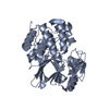


 PDBj
PDBj


 Assembly


 Mass: 54.938 Da / Num. of mol.: 8 / Source method: obtained synthetically / Formula: Mn
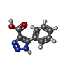
 Mass: 189.171 Da / Num. of mol.: 4 / Source method: obtained synthetically / Formula: C9H7N3O2
 Mass: 18.015 Da / Num. of mol.: 358 / Source method: isolated from a natural source / Formula: H2O
 Sample preparation
 Processing