| Entry | Database: PDB / ID: 5ugu
|
|---|
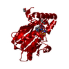
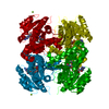
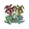
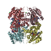
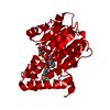
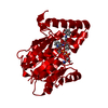
| Title | Crystal structure of M. tuberculosis InhA inhibited by PT506 |
|---|
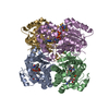
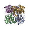
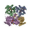
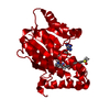
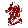
 Components Components | Enoyl-[acyl-carrier-protein] reductase [NADH] |
|---|
 Keywords Keywords | OXIDOREDUCTASE / bacterial enoyl-ACP reductase / diphenylether / residence time |
|---|
| Function / homology |  Function and homology information Function and homology information
trans-2-enoyl-CoA reductase (NADH) activity / mycolic acid biosynthetic process / enoyl-[acyl-carrier-protein] reductase (NADH) / fatty acid elongation / enoyl-[acyl-carrier-protein] reductase (NADH) activity / NAD+ binding / peptidoglycan-based cell wall / fatty acid binding / response to antibiotic / plasma membraneSimilarity search - Function : / Enoyl-[acyl-carrier-protein] reductase (NADH) / Enoyl-(Acyl carrier protein) reductase / Short-chain dehydrogenase/reductase SDR / NAD(P)-binding Rossmann-like Domain / NAD(P)-binding domain superfamily / Rossmann fold / 3-Layer(aba) Sandwich / Alpha BetaSimilarity search - Domain/homology |
|---|
| Biological species |   Mycobacterium tuberculosis (bacteria) Mycobacterium tuberculosis (bacteria) |
|---|
| Method |  X-RAY DIFFRACTION / SYNCHROTRON / MOLECULAR REPLACEMENT / Resolution: 1.95 Å X-RAY DIFFRACTION / SYNCHROTRON / MOLECULAR REPLACEMENT / Resolution: 1.95 Å |
|---|
 Authors Authors | Eltschkner, S. / Pschibul, A. / Spagnuolo, L.A. / Yu, W. / Tonge, P.J. / Kisker, C. |
|---|
| Funding support |  Germany, 1items Germany, 1items | Organization | Grant number | Country |
|---|
| German Research Foundation | SFB 630 | Germany |
|
|---|
 Citation Citation | Journal: J. Am. Chem. Soc. / Year: 2017
Title: Evaluating the Contribution of Transition-State Destabilization to Changes in the Residence Time of Triazole-Based InhA Inhibitors.
Authors: Spagnuolo, L.A. / Eltschkner, S. / Yu, W. / Daryaee, F. / Davoodi, S. / Knudson, S.E. / Allen, E.K. / Merino, J. / Pschibul, A. / Moree, B. / Thivalapill, N. / Truglio, J.J. / Salafsky, J. / ...Authors: Spagnuolo, L.A. / Eltschkner, S. / Yu, W. / Daryaee, F. / Davoodi, S. / Knudson, S.E. / Allen, E.K. / Merino, J. / Pschibul, A. / Moree, B. / Thivalapill, N. / Truglio, J.J. / Salafsky, J. / Slayden, R.A. / Kisker, C. / Tonge, P.J. |
|---|
| History | | Deposition | Jan 10, 2017 | Deposition site: RCSB / Processing site: PDBE |
|---|
| Revision 1.0 | Feb 15, 2017 | Provider: repository / Type: Initial release |
|---|
| Revision 1.1 | Mar 22, 2017 | Group: Database references |
|---|
| Revision 1.2 | Sep 6, 2017 | Group: Author supporting evidence / Category: pdbx_audit_support / Item: _pdbx_audit_support.funding_organization |
|---|
| Revision 1.3 | Jan 17, 2024 | Group: Data collection / Database references / Refinement description
Category: chem_comp_atom / chem_comp_bond ...chem_comp_atom / chem_comp_bond / database_2 / pdbx_initial_refinement_model
Item: _database_2.pdbx_DOI / _database_2.pdbx_database_accession |
|---|
|
|---|
 Movie
Movie Controller


 Open data
 Basic information
 Structure visualization
 Downloads & links
 Other downloads
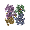


 PDBj
PDBj

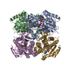
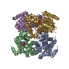
 Assembly


 Mass: 663.425 Da / Num. of mol.: 1 / Source method: obtained synthetically / Formula: C21H27N7O14P2 / Comment: NAD*YM
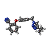
 Mass: 332.356 Da / Num. of mol.: 1 / Source method: obtained synthetically / Formula: C19H16N4O2
 Mass: 18.015 Da / Num. of mol.: 174 / Source method: isolated from a natural source / Formula: H2O
 Sample preparation
 Processing