 Movie
Movie Controller

[English] 日本語
 Yorodumi
- PDB-5ubb: Crystal structure of human alpha N-terminal protein methyltransfe... -
+ Open data

- Basic information
| Entry | Database: PDB / ID: 5ubb | ||||||
|---|---|---|---|---|---|---|---|
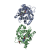
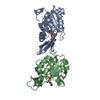
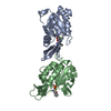
| Title | Crystal structure of human alpha N-terminal protein methyltransferase 1B | ||||||
 Components Components | Alpha N-terminal protein methyltransferase 1B | ||||||
 Keywords Keywords | TRANSFERASE / methyl transferase / Structural Genomics / Structural Genomics Consortium / SGC | ||||||
| Function / homology |  Function and homology information Function and homology informationprotein N-terminal monomethyltransferase / N-terminal protein amino acid methylation / N-terminal protein N-methyltransferase activity / nucleus / cytoplasm Similarity search - Function | ||||||
| Biological species |  Homo sapiens (human) Homo sapiens (human) | ||||||
| Method |  X-RAY DIFFRACTION / MOLECULAR REPLACEMENT / Resolution: 2 Å X-RAY DIFFRACTION / MOLECULAR REPLACEMENT / Resolution: 2 Å | ||||||
 Authors Authors | Dong, C. / Zhu, L. / Tempel, W. / Dong, A. / Bountra, C. / Arrowsmith, C.H. / Edwards, A.M. / Min, J. / Structural Genomics Consortium (SGC) | ||||||
 Citation Citation | Journal: Commun Biol / Year: 2018 Title: An asparagine/glycine switch governs product specificity of human N-terminal methyltransferase NTMT2. Authors: Dong, C. / Dong, G. / Li, L. / Zhu, L. / Tempel, W. / Liu, Y. / Huang, R. / Min, J. | ||||||
| History |
|
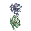
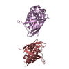
- Structure visualization
| Structure viewer | Molecule: MolmilJmol/JSmol |
|---|
- Downloads & links
-Download
| PDBx/mmCIF format | 5ubb.cif.gz | 60.5 KB | Display | PDBx/mmCIF format |
|---|---|---|---|---|
| PDB format | pdb5ubb.ent.gz | 41.7 KB | Display | PDB format |
| PDBx/mmJSON format | 5ubb.json.gz | Tree view | PDBx/mmJSON format | |
| Others |  Other downloads Other downloads |
-Validation report
| Arichive directory | https://data.pdbj.org/pub/pdb/validation_reports/ub/5ubbftp://data.pdbj.org/pub/pdb/validation_reports/ub/5ubb | HTTPS FTP |
|---|
-Related structure data


-Links
 PDBj
PDBj
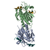
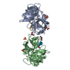
- Assembly
| Deposited unit | 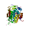
| ||||||||
|---|---|---|---|---|---|---|---|---|---|
| 1 | 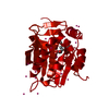
| ||||||||
| Unit cell |
| ||||||||
| Components on special symmetry positions |
|
-Components
| #1: Protein | Mass: 25005.688 Da / Num. of mol.: 1 / Fragment: UNP residues 58-278 Source method: isolated from a genetically manipulated source Source: (gene. exp.) Homo sapiens (human) / Gene: METTL11B, C1orf184, NRMT2 / Plasmid: pET28-MKH8SUMO / Production host:  References: UniProt: Q5VVY1, protein N-terminal monomethyltransferase | ||
|---|---|---|---|
| #2: Chemical | ChemComp-SAM /   Mass: 398.437 Da / Num. of mol.: 1 / Source method: obtained synthetically / Formula: C15H22N6O5S Mass: 398.437 Da / Num. of mol.: 1 / Source method: obtained synthetically / Formula: C15H22N6O5S | ||
| #3: Chemical | ChemComp-UNX /   Num. of mol.: 26 / Source method: obtained synthetically Num. of mol.: 26 / Source method: obtained synthetically#4: Water | ChemComp-HOH / |  Mass: 18.015 Da / Num. of mol.: 63 / Source method: isolated from a natural source / Formula: H2O Mass: 18.015 Da / Num. of mol.: 63 / Source method: isolated from a natural source / Formula: H2O |
-Experimental details
-Experiment
| Experiment | Method: X-RAY DIFFRACTION / Number of used crystals: 1 |
|---|
- Sample preparation
| Crystal | Density Matthews: 2.56 Å3/Da / Density % sol: 52.02 % |
|---|---|
| Crystal grow | Temperature: 291 K / Method: vapor diffusion, sitting drop / Details: 20% PEG3350, 0.2 M sodium acetate |
-Data collection
| Diffraction | Mean temperature: 100 K | ||||||||||||||||||||||||||||||
|---|---|---|---|---|---|---|---|---|---|---|---|---|---|---|---|---|---|---|---|---|---|---|---|---|---|---|---|---|---|---|---|
| Diffraction source | Source: ROTATING ANODE / Type: RIGAKU FR-E SUPERBRIGHT / Wavelength: 1.5418 Å | ||||||||||||||||||||||||||||||
| Detector | Type: RIGAKU SATURN A200 / Detector: CCD / Date: Aug 25, 2016 | ||||||||||||||||||||||||||||||
| Radiation | Protocol: SINGLE WAVELENGTH / Monochromatic (M) / Laue (L): M / Scattering type: x-ray | ||||||||||||||||||||||||||||||
| Radiation wavelength | Wavelength: 1.5418 Å / Relative weight: 1 | ||||||||||||||||||||||||||||||
| Reflection | Resolution: 2→19.98 Å / Num. obs: 16827 / % possible obs: 94.4 % / Redundancy: 2.6 % / Biso Wilson estimate: 25.74 Å2 / CC1/2: 0.996 / Rmerge(I) obs: 0.086 / Rpim(I) all: 0.06 / Rrim(I) all: 0.106 / Net I/σ(I): 10.2 / Num. measured all: 44067 / Scaling rejects: 0 | ||||||||||||||||||||||||||||||
| Reflection shell | Diffraction-ID: 1
|
- Processing
| Software |
| ||||||||||||||||||||||||||||||||||||||||||||||||||||||||||||||||||||||||||||||||||||||||||||||||||||||||||||
|---|---|---|---|---|---|---|---|---|---|---|---|---|---|---|---|---|---|---|---|---|---|---|---|---|---|---|---|---|---|---|---|---|---|---|---|---|---|---|---|---|---|---|---|---|---|---|---|---|---|---|---|---|---|---|---|---|---|---|---|---|---|---|---|---|---|---|---|---|---|---|---|---|---|---|---|---|---|---|---|---|---|---|---|---|---|---|---|---|---|---|---|---|---|---|---|---|---|---|---|---|---|---|---|---|---|---|---|---|---|
| Refinement | Method to determine structure: MOLECULAR REPLACEMENT / Resolution: 2→18.98 Å / Cor.coef. Fo:Fc: 0.918 / Cor.coef. Fo:Fc free: 0.893 / SU R Cruickshank DPI: 0.182 / Cross valid method: FREE R-VALUE / σ(F): 0 / SU R Blow DPI: 0.187 / SU Rfree Blow DPI: 0.168 / SU Rfree Cruickshank DPI: 0.166 Details: Intensities of only approx. 64 degrees of the data collection sweep were merged and used for model refinement. Subsequent diffraction images show ice rings, possibly due to malfunctioning ...Details: Intensities of only approx. 64 degrees of the data collection sweep were merged and used for model refinement. Subsequent diffraction images show ice rings, possibly due to malfunctioning crystal cooling. The protein was crystallized in the presence of a putative inhibitor, but electron density maps did not fully resolve the expected inhibitor.
| ||||||||||||||||||||||||||||||||||||||||||||||||||||||||||||||||||||||||||||||||||||||||||||||||||||||||||||
| Displacement parameters | Biso max: 73.45 Å2 / Biso mean: 27.38 Å2 / Biso min: 11.65 Å2
| ||||||||||||||||||||||||||||||||||||||||||||||||||||||||||||||||||||||||||||||||||||||||||||||||||||||||||||
| Refine analyze | Luzzati coordinate error obs: 0.3 Å | ||||||||||||||||||||||||||||||||||||||||||||||||||||||||||||||||||||||||||||||||||||||||||||||||||||||||||||
| Refinement step | Cycle: final / Resolution: 2→18.98 Å
| ||||||||||||||||||||||||||||||||||||||||||||||||||||||||||||||||||||||||||||||||||||||||||||||||||||||||||||
| Refine LS restraints |
| ||||||||||||||||||||||||||||||||||||||||||||||||||||||||||||||||||||||||||||||||||||||||||||||||||||||||||||
| LS refinement shell | Resolution: 2→2.14 Å / Total num. of bins used: 8
|
