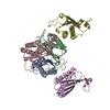
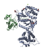
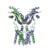
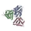
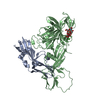
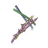
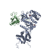
Entry Database : PDB / ID : 5u7gTitle Crystal Structure of the Catalytic Core of CBP CREB-binding protein Keywords / / / / Function / homology Function Domain/homology Component
/ / / / / / / / / / / / / / / / / / / / / / / / / / / / / / / / / / / / / / / / / / / / / / / / / / / / / / / / / / / / / / / / / / / / / / / / / / / / / / / / / / / / / / / / / / / / / / / / / / / / / / / / / / / / / / / / / / / / / / / / / / / / / / / / / / / / / / / / / / / / / / / / / / / Biological species Mus musculus (house mouse)Method / / / Resolution : 2.401 Å Authors Park, S. / Stanfield, R.L. / Martinez-Yamout, M.M. / Dyson, H.J. / Wilson, I.A. / Wright, P.E. Funding support Organization Grant number Country National Institutes of Health/National Cancer Institute (NIH/NCI) CA096865 National Institutes of Health/National Cancer Institute (NIH/NCI) Y1-CO-1020 National Institutes of Health/National Institute of General Medical Sciences (NIH/NIGMS) Y1-GM-1104
Journal : Proc. Natl. Acad. Sci. U.S.A. / Year : 2017Title : Role of the CBP catalytic core in intramolecular SUMOylation and control of histone H3 acetylation.Authors : Park, S. / Stanfield, R.L. / Martinez-Yamout, M.A. / Dyson, H.J. / Wilson, I.A. / Wright, P.E. History Deposition Dec 12, 2016 Deposition site / Processing site Revision 1.0 Jun 21, 2017 Provider / Type Revision 1.1 Jul 5, 2017 Group / Category Item _citation.country / _citation.journal_abbrev ... _citation.country / _citation.journal_abbrev / _citation.journal_id_ASTM / _citation.journal_id_CSD / _citation.pdbx_database_id_PubMed / _citation.title Revision 1.2 Jul 12, 2017 Group / Category Item / _citation.page_first / _citation.page_lastRevision 1.3 Sep 27, 2017 Group / Category / Item Revision 1.4 Dec 4, 2019 Group / Category / Item Revision 1.5 Oct 4, 2023 Group / Database references / Refinement descriptionCategory chem_comp_atom / chem_comp_bond ... chem_comp_atom / chem_comp_bond / database_2 / pdbx_initial_refinement_model Item / _database_2.pdbx_database_accession
Show all Show less
 Movie
Movie Controller


 Open data
 Basic information
 Components
 Keywords
 Function and homology information
 X-RAY DIFFRACTION /
  Authors
 United States, 3items
  Citation
 Structure visualization
 Downloads & links
 Other downloads
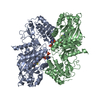

 PDBj
PDBj


 Assembly


 Mass: 65.409 Da / Num. of mol.: 8
 Mass: 18.015 Da / Num. of mol.: 45 / Source method: isolated from a natural source / Formula: H2O
 Sample preparation
 Processing