 Movie
Movie Controller


+ Open data

- Basic information
| Entry | Database: PDB / ID: 5tzb | ||||||
|---|---|---|---|---|---|---|---|
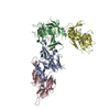
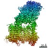
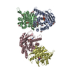
| Title | Burkholderia sp. beta-aminopeptidase | ||||||
 Components Components | D-aminopeptidase | ||||||
 Keywords Keywords | HYDROLASE / beta-aminopeptidase / enzyme / Burkholderia / Ntn hydrolases / beta-amino acids | ||||||
| Function / homology | L-amino peptidase D-ALA esterase/amidase / L-amino peptidase D-ALA esterase/amidase / 4-Layer Sandwich / Alpha Beta / :  Function and homology information Function and homology information | ||||||
| Biological species |  Burkholderia sp. LK4 (bacteria) Burkholderia sp. LK4 (bacteria) | ||||||
| Method |  X-RAY DIFFRACTION / SYNCHROTRON / MOLECULAR REPLACEMENT / Resolution: 1.977 Å X-RAY DIFFRACTION / SYNCHROTRON / MOLECULAR REPLACEMENT / Resolution: 1.977 Å | ||||||
 Authors Authors | McGowan, S. / Drinkwater, N. / John, M. / Dumsday, G. | ||||||
 Citation Citation | Journal: Acta Crystallogr F Struct Biol Commun / Year: 2017 Title: Crystal structure of a beta-aminopeptidase from an Australian Burkholderia sp. Authors: John-White, M. / Dumsday, G.J. / Johanesen, P. / Lyras, D. / Drinkwater, N. / McGowan, S. | ||||||
| History |
|
- Structure visualization
| Structure viewer | Molecule: MolmilJmol/JSmol |
|---|
- Downloads & links
-Download
| PDBx/mmCIF format | 5tzb.cif.gz | 1 MB | Display | PDBx/mmCIF format |
|---|---|---|---|---|
| PDB format | pdb5tzb.ent.gz | 888 KB | Display | PDB format |
| PDBx/mmJSON format | 5tzb.json.gz | Tree view | PDBx/mmJSON format | |
| Others |  Other downloads Other downloads |
-Validation report
| Arichive directory | https://data.pdbj.org/pub/pdb/validation_reports/tz/5tzbftp://data.pdbj.org/pub/pdb/validation_reports/tz/5tzb | HTTPS FTP |
|---|
-Related structure data
| Related structure data | 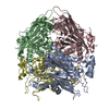 1b65S S: Starting model for refinement |
|---|---|
| Similar structure data |


-Links
 PDBj
PDBj- Assembly
| Deposited unit | 
| ||||||||
|---|---|---|---|---|---|---|---|---|---|
| 1 | 
| ||||||||
| 2 | 
| ||||||||
| Unit cell |
|
-Components
| #1: Protein | Mass: 40087.938 Da / Num. of mol.: 8 Source method: isolated from a genetically manipulated source Source: (gene. exp.) Burkholderia sp. LK4 (bacteria) / Gene: VK92_01140 / Production host: #2: Chemical | ChemComp-CA /   Mass: 40.078 Da / Num. of mol.: 4 Mass: 40.078 Da / Num. of mol.: 4Source method: isolated from a genetically manipulated source Formula: Ca #3: Water | ChemComp-HOH / |  Mass: 18.015 Da / Num. of mol.: 1782 / Source method: isolated from a natural source / Formula: H2O Mass: 18.015 Da / Num. of mol.: 1782 / Source method: isolated from a natural source / Formula: H2O |
|---|
-Experimental details
-Experiment
| Experiment | Method: X-RAY DIFFRACTION / Number of used crystals: 1 |
|---|
- Sample preparation
| Crystal | Density Matthews: 2.22 Å3/Da / Density % sol: 44.68 % |
|---|---|
| Crystal grow | Temperature: 293 K / Method: vapor diffusion, hanging drop / pH: 7 Details: 35 % 2-methyl-2,4-pentanediol, 0.1 M sodium cacodylate pH 7.0 and 5 % PEG8000 |
-Data collection
| Diffraction | Mean temperature: 100 K | ||||||||||||||||||
|---|---|---|---|---|---|---|---|---|---|---|---|---|---|---|---|---|---|---|---|
| Diffraction source | Source: SYNCHROTRON / Site: Australian Synchrotron  / Beamline: MX1 / Wavelength: 0.95369 Å / Beamline: MX1 / Wavelength: 0.95369 Å | ||||||||||||||||||
| Detector | Type: ADSC QUANTUM 210r / Detector: CCD / Date: Dec 10, 2015 | ||||||||||||||||||
| Radiation | Monochromator: Double-crystal Si(111) water-cooled / Protocol: SINGLE WAVELENGTH / Monochromatic (M) / Laue (L): M / Scattering type: x-ray | ||||||||||||||||||
| Radiation wavelength | Wavelength: 0.95369 Å / Relative weight: 1 | ||||||||||||||||||
| Reflection | Resolution: 1.98→45.711 Å / Num. obs: 190171 / % possible obs: 97.4 % / Redundancy: 4.1 % / Biso Wilson estimate: 16.5 Å2 / CC1/2: 0.986 / Rmerge(I) obs: 0.14 / Net I/σ(I): 8.1 | ||||||||||||||||||
| Reflection shell |
|
- Processing
| Software |
| |||||||||||||||||||||||||||||||||||||||||||||||||||||||||||||||||||||||||||||||||||||||||||||||||||||||||||||||||||||||||||||||||||||||||||||||||||||||||||||||||||||||||||||||||||||||||||||||||||||||||||||||||||||||||
|---|---|---|---|---|---|---|---|---|---|---|---|---|---|---|---|---|---|---|---|---|---|---|---|---|---|---|---|---|---|---|---|---|---|---|---|---|---|---|---|---|---|---|---|---|---|---|---|---|---|---|---|---|---|---|---|---|---|---|---|---|---|---|---|---|---|---|---|---|---|---|---|---|---|---|---|---|---|---|---|---|---|---|---|---|---|---|---|---|---|---|---|---|---|---|---|---|---|---|---|---|---|---|---|---|---|---|---|---|---|---|---|---|---|---|---|---|---|---|---|---|---|---|---|---|---|---|---|---|---|---|---|---|---|---|---|---|---|---|---|---|---|---|---|---|---|---|---|---|---|---|---|---|---|---|---|---|---|---|---|---|---|---|---|---|---|---|---|---|---|---|---|---|---|---|---|---|---|---|---|---|---|---|---|---|---|---|---|---|---|---|---|---|---|---|---|---|---|---|---|---|---|---|---|---|---|---|---|---|---|---|---|---|---|---|---|---|---|---|
| Refinement | Method to determine structure: MOLECULAR REPLACEMENT Starting model: 1B65 Resolution: 1.977→45.711 Å / SU ML: 0.24 / Cross valid method: FREE R-VALUE / σ(F): 1.33 / Phase error: 23.76 / Stereochemistry target values: ML
| |||||||||||||||||||||||||||||||||||||||||||||||||||||||||||||||||||||||||||||||||||||||||||||||||||||||||||||||||||||||||||||||||||||||||||||||||||||||||||||||||||||||||||||||||||||||||||||||||||||||||||||||||||||||||
| Solvent computation | Shrinkage radii: 0.9 Å / VDW probe radii: 1.11 Å / Solvent model: FLAT BULK SOLVENT MODEL | |||||||||||||||||||||||||||||||||||||||||||||||||||||||||||||||||||||||||||||||||||||||||||||||||||||||||||||||||||||||||||||||||||||||||||||||||||||||||||||||||||||||||||||||||||||||||||||||||||||||||||||||||||||||||
| Refinement step | Cycle: LAST / Resolution: 1.977→45.711 Å
| |||||||||||||||||||||||||||||||||||||||||||||||||||||||||||||||||||||||||||||||||||||||||||||||||||||||||||||||||||||||||||||||||||||||||||||||||||||||||||||||||||||||||||||||||||||||||||||||||||||||||||||||||||||||||
| Refine LS restraints |
| |||||||||||||||||||||||||||||||||||||||||||||||||||||||||||||||||||||||||||||||||||||||||||||||||||||||||||||||||||||||||||||||||||||||||||||||||||||||||||||||||||||||||||||||||||||||||||||||||||||||||||||||||||||||||
| LS refinement shell |
| |||||||||||||||||||||||||||||||||||||||||||||||||||||||||||||||||||||||||||||||||||||||||||||||||||||||||||||||||||||||||||||||||||||||||||||||||||||||||||||||||||||||||||||||||||||||||||||||||||||||||||||||||||||||||
| Refinement TLS params. | Method: refined / Origin x: 23.6775 Å / Origin y: -38.2948 Å / Origin z: 13.3942 Å
| |||||||||||||||||||||||||||||||||||||||||||||||||||||||||||||||||||||||||||||||||||||||||||||||||||||||||||||||||||||||||||||||||||||||||||||||||||||||||||||||||||||||||||||||||||||||||||||||||||||||||||||||||||||||||
| Refinement TLS group | Selection details: ALL |
