 Movie
Movie Controller


+ Open data

- Basic information
| Entry | Database: PDB / ID: 5tuc | ||||||
|---|---|---|---|---|---|---|---|
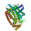
| Title | Crystal Structure of the Sus TBC1D15 GAP Domain | ||||||
 Components Components | Sus TBC1D15 GAP Domain | ||||||
 Keywords Keywords | HYDROLASE ACTIVATOR / Sus (Sus scrofa) / TBC1D15 / GAP (GTPase-activating Protein) / GTPase / PROTEIN BINDING | ||||||
| Function / homology | Small G protein signalling modulator 1/2, Rab-binding domain / Rab-binding domain (RBD) / Domain in Tre-2, BUB2p, and Cdc16p. Probable Rab-GAPs. / Rab-GTPase-TBC domain / Rab-GTPase-TBC domain superfamily / Rab-GTPase-TBC domain / TBC/rab GAP domain profile. / cytoplasm / TBC1 domain family member 15 Function and homology information Function and homology information | ||||||
| Biological species |  | ||||||
| Method |  X-RAY DIFFRACTION / SYNCHROTRON / MOLECULAR REPLACEMENT / Resolution: 2.5 Å X-RAY DIFFRACTION / SYNCHROTRON / MOLECULAR REPLACEMENT / Resolution: 2.5 Å | ||||||
 Authors Authors | Chen, Y.-N. / Wang, W. / Cheng, D. / Ge, Y. / Gu, X. / Zhou, X.E. / Ye, F. / Xu, H.E. / Lv, Z. | ||||||
 Citation Citation | Journal: Protein Sci. / Year: 2017 Title: Crystal structure of TBC1D15 GTPase-activating protein (GAP) domain and its activity on Rab GTPases. Authors: Chen, Y.N. / Gu, X. / Zhou, X.E. / Wang, W. / Cheng, D. / Ge, Y. / Ye, F. / Xu, H.E. / Lv, Z. | ||||||
| History |
|
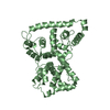
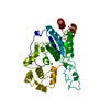
- Structure visualization
| Structure viewer | Molecule: MolmilJmol/JSmol |
|---|
- Downloads & links
-Download
| PDBx/mmCIF format | 5tuc.cif.gz | 147 KB | Display | PDBx/mmCIF format |
|---|---|---|---|---|
| PDB format | pdb5tuc.ent.gz | 116.3 KB | Display | PDB format |
| PDBx/mmJSON format | 5tuc.json.gz | Tree view | PDBx/mmJSON format | |
| Others |  Other downloads Other downloads |
-Validation report
| Arichive directory | https://data.pdbj.org/pub/pdb/validation_reports/tu/5tucftp://data.pdbj.org/pub/pdb/validation_reports/tu/5tuc | HTTPS FTP |
|---|
-Related structure data
| Related structure data | 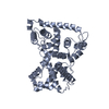 5tubC  2qfzS C: citing same article ( S: Starting model for refinement |
|---|---|
| Similar structure data |


-Links
 PDBj
PDBj- Assembly
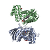
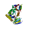
| Deposited unit | 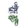
| ||||||||
|---|---|---|---|---|---|---|---|---|---|
| 1 | 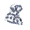
| ||||||||
| 2 | 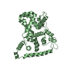
| ||||||||
| Unit cell |
| ||||||||
| Components on special symmetry positions |
|
-Components
| #1: Protein | Mass: 41557.730 Da / Num. of mol.: 2 / Fragment: UNP residues 270-617 Source method: isolated from a genetically manipulated source Source: (gene. exp.)  #2: Water | ChemComp-HOH / |  Mass: 18.015 Da / Num. of mol.: 154 / Source method: isolated from a natural source / Formula: H2O Mass: 18.015 Da / Num. of mol.: 154 / Source method: isolated from a natural source / Formula: H2O |
|---|
-Experimental details
-Experiment
| Experiment | Method: X-RAY DIFFRACTION / Number of used crystals: 1 |
|---|
- Sample preparation
| Crystal | Density Matthews: 2.94 Å3/Da / Density % sol: 58.16 % |
|---|---|
| Crystal grow | Temperature: 293 K / Method: vapor diffusion / pH: 6.5 Details: 50 mM MES sodium Salt (pH 6.5), 20% (w/v) PEG1000, 100 mM sodium chloride and 200 mM magnesium chloride PH range: 6.5 |
-Data collection
| Diffraction | Mean temperature: 100 K |
|---|---|
| Diffraction source | Source: SYNCHROTRON / Site: SSRF  / Beamline: BL17U / Wavelength: 1 Å / Beamline: BL17U / Wavelength: 1 Å |
| Detector | Type: MARRESEARCH / Detector: CCD / Date: Sep 26, 2015 |
| Radiation | Protocol: SINGLE WAVELENGTH / Monochromatic (M) / Laue (L): M / Scattering type: x-ray |
| Radiation wavelength | Wavelength: 1 Å / Relative weight: 1 |
| Reflection | Resolution: 2.5→50 Å / Num. obs: 35232 / % possible obs: 100 % / Redundancy: 39.6 % / Rmerge(I) obs: 0.172 / Net I/σ(I): 22.2 |
| Reflection shell | Resolution: 2.5→2.61 Å / Rmerge(I) obs: 1.254 / Mean I/σ(I) obs: 4.4 |
- Processing
| Software |
| |||||||||||||||||||||||||||||||||||||||||||||||||||||||||||||||||||||||||||||||||||||||||||||||||||||||||||||||||||||||||||||||||||||
|---|---|---|---|---|---|---|---|---|---|---|---|---|---|---|---|---|---|---|---|---|---|---|---|---|---|---|---|---|---|---|---|---|---|---|---|---|---|---|---|---|---|---|---|---|---|---|---|---|---|---|---|---|---|---|---|---|---|---|---|---|---|---|---|---|---|---|---|---|---|---|---|---|---|---|---|---|---|---|---|---|---|---|---|---|---|---|---|---|---|---|---|---|---|---|---|---|---|---|---|---|---|---|---|---|---|---|---|---|---|---|---|---|---|---|---|---|---|---|---|---|---|---|---|---|---|---|---|---|---|---|---|---|---|---|
| Refinement | Method to determine structure: MOLECULAR REPLACEMENT Starting model: 2QFZ Resolution: 2.5→87.62 Å / SU ML: 0.3 / Cross valid method: FREE R-VALUE / σ(F): 1.97 / Phase error: 25.69 / Stereochemistry target values: ML
| |||||||||||||||||||||||||||||||||||||||||||||||||||||||||||||||||||||||||||||||||||||||||||||||||||||||||||||||||||||||||||||||||||||
| Solvent computation | Shrinkage radii: 0.7 Å / VDW probe radii: 1 Å / Solvent model: FLAT BULK SOLVENT MODEL | |||||||||||||||||||||||||||||||||||||||||||||||||||||||||||||||||||||||||||||||||||||||||||||||||||||||||||||||||||||||||||||||||||||
| Refinement step | Cycle: LAST / Resolution: 2.5→87.62 Å
| |||||||||||||||||||||||||||||||||||||||||||||||||||||||||||||||||||||||||||||||||||||||||||||||||||||||||||||||||||||||||||||||||||||
| Refine LS restraints |
| |||||||||||||||||||||||||||||||||||||||||||||||||||||||||||||||||||||||||||||||||||||||||||||||||||||||||||||||||||||||||||||||||||||
| LS refinement shell |
|
