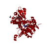
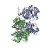
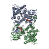
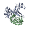
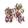
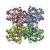
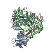
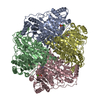
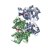
[English] 日本語
Yorodumi
- PDB-5tow: Crystal structure of the inactive form of S-adenosyl-L-homocystei... -

+
Open data


ID or keywords:

Loading...

-
Basic information

Entry
Database: PDB / ID: 5tow
TitleCrystal structure of the inactive form of S-adenosyl-L-homocysteine hydrolase from Thermotoga maritima in ternary complex with NADH and Adenosine
ComponentsAdenosylhomocysteinase
KeywordsHYDROLASE / REGULATION OF SAM-DEPENDENT METHYLATION REACTIONS
Function / homology
Function and homology information


adenosylhomocysteinase / adenosylhomocysteinase activity / S-adenosylmethionine cycle / one-carbon metabolic process / cytosol
Similarity search - Function
Adenosylhomocysteinase-like / Adenosylhomocysteinase-like / S-adenosyl-L-homocysteine hydrolase, conserved site / Adenosylhomocysteinase-like superfamily / S-adenosyl-L-homocysteine hydrolase, NAD binding domain / S-adenosyl-L-homocysteine hydrolase / S-adenosyl-L-homocysteine hydrolase signature 1. / S-adenosyl-L-homocysteine hydrolase signature 2. / S-adenosyl-L-homocysteine hydrolase / S-adenosyl-L-homocysteine hydrolase, NAD binding domain ...Adenosylhomocysteinase-like / Adenosylhomocysteinase-like / S-adenosyl-L-homocysteine hydrolase, conserved site / Adenosylhomocysteinase-like superfamily / S-adenosyl-L-homocysteine hydrolase, NAD binding domain / S-adenosyl-L-homocysteine hydrolase / S-adenosyl-L-homocysteine hydrolase signature 1. / S-adenosyl-L-homocysteine hydrolase signature 2. / S-adenosyl-L-homocysteine hydrolase / S-adenosyl-L-homocysteine hydrolase, NAD binding domain / S-adenosyl-L-homocysteine hydrolase, NAD binding domain / NAD(P)-binding Rossmann-like Domain / NAD(P)-binding domain superfamily / Rossmann fold / 3-Layer(aba) Sandwich / Alpha Beta
Similarity search - Domain/homology
ADENOSINE / 1,4-DIHYDRONICOTINAMIDE ADENINE DINUCLEOTIDE / Adenosylhomocysteinase
Similarity search - Component
Biological speciesThermotoga maritima (bacteria)
MethodX-RAY DIFFRACTION / SYNCHROTRON / MOLECULAR REPLACEMENT / Resolution: 1.75 Å
AuthorsCzyrko, J. / Brzezinski, K.
Funding support Poland, 1items
OrganizationGrant numberCountry
National Science Centre, PolandUMO-2013/09/B/NZ1/01880 Poland
CitationJournal: Int. J. Biol. Macromol. / Year: 2017
Title: S-adenosyl-L-homocysteine hydrolase from a hyperthermophile (Thermotoga maritima) is expressed in Escherichia coli in inactive form - Biochemical and structural studies.
Authors: Brzezinski, K. / Czyrko, J. / Sliwiak, J. / Nalewajko-Sieliwoniuk, E. / Jaskolski, M. / Nocek, B. / Dauter, Z.
History
DepositionOct 19, 2016Deposition site: RCSB / Processing site: PDBE
Revision 1.0Jul 5, 2017Provider: repository / Type: Initial release
Revision 1.1Apr 22, 2020Group: Database references / Category: pdbx_related_exp_data_set
Revision 1.2Jan 17, 2024Group: Data collection / Database references / Refinement description
Category: chem_comp_atom / chem_comp_bond ...chem_comp_atom / chem_comp_bond / database_2 / pdbx_initial_refinement_model
Item: _database_2.pdbx_DOI / _database_2.pdbx_database_accession

-
Structure visualization

Structure viewerMolecule:
MolmilJmol/JSmol

Downloads & links

-
Assembly

Deposited unit
A: Adenosylhomocysteinase
B: Adenosylhomocysteinase
hetero molecules


Theoretical massNumber of molelcules
Total (without water)91,4797
Polymers90,3582
Non-polymers1,1225
Water10,467581
1
A: Adenosylhomocysteinase
B: Adenosylhomocysteinase
hetero molecules

A: Adenosylhomocysteinase
B: Adenosylhomocysteinase
hetero molecules


Theoretical massNumber of molelcules
Total (without water)182,95914
Polymers180,7154
Non-polymers2,24410
Water724
TypeNameSymmetry operationNumber
identity operation1_555x,y,z1
crystal symmetry operation2_656-x+1,y,-z+11
Buried area21060 Å2
ΔGint-178 kcal/mol
Surface area56150 Å2
MethodPISA
Unit cell
Length a, b, c (Å)120.381, 105.509, 85.535
Angle α, β, γ (deg.)90.00, 108.76, 90.00
Int Tables number5
Space group name H-MC121

-
Components

-
Protein , 1 types, 2 molecules AB

#1: Protein Adenosylhomocysteinase / S-adenosyl-L-homocysteine hydrolase / AdoHcyase


Mass: 45178.848 Da / Num. of mol.: 2
Source method: isolated from a genetically manipulated source
Details: ...Details: SNAMNTGEMKINWVSRYMPLLNKIAEEYSREKPLSGFTVGMSIHLEAKTAYLAITLSKLGAKVVITGSNPLSTQDDVAEALRSKGITVYARRTHDESIYRENLMKVLDERPDFIIDDGGDLTVISHTEREEVLENLKGVSEETTTGVRRLKALEETGKLRVPVIAVNDSKMKYLFDNRYGTGQSTWDAIMRNTNLLVAGKNVVVAGYGWCGRGIALRAAGLGARVIVTEVDPVKAVEAIMDGFTVMPMKEAVKIADFVITASGNTDVLSKEDILSLKDGAVLANAGHFNVEIPVRVLEEIAVEKFEARPNVTGYTLENGKTVFLLAEGRLVNLAAGDGHPVEIMDLSFALQIFAVLYLLENHRKMSPKVYMLPDEIDERVARMKLDSLGVKIDELTEKQRRYLRSWQ
Source: (gene. exp.) Thermotoga maritima (strain ATCC 43589 / MSB8 / DSM 3109 / JCM 10099) (bacteria)
Strain: ATCC 43589 / MSB8 / DSM 3109 / JCM 10099 / Gene: ahcY, TM_0172 / Plasmid: pMCSG9 / Details (production host): pMCSG9 / Production host: Escherichia coli (E. coli) / Strain (production host): BL21 / Variant (production host): CodonPlus RIPL / References: UniProt: O51933, adenosylhomocysteinase

-
Non-polymers , 5 types, 586 molecules

#2: Chemical ChemComp-ADN / ADENOSINE


Mass: 267.241 Da / Num. of mol.: 1 / Source method: obtained synthetically / Formula: C10H13N5O4
#3: Chemical ChemComp-MPD / (4S)-2-METHYL-2,4-PENTANEDIOL


Mass: 118.174 Da / Num. of mol.: 1 / Source method: obtained synthetically / Formula: C6H14O2 / Comment: precipitant*YM
#4: Chemical ChemComp-NAI / 1,4-DIHYDRONICOTINAMIDE ADENINE DINUCLEOTIDE / NADH


Mass: 665.441 Da / Num. of mol.: 1 / Source method: obtained synthetically / Formula: C21H29N7O14P2
#5: Chemical ChemComp-CL / CHLORIDE ION


Mass: 35.453 Da / Num. of mol.: 2 / Source method: obtained synthetically / Formula: Cl
#6: Water ChemComp-HOH / water


Mass: 18.015 Da / Num. of mol.: 581 / Source method: isolated from a natural source / Formula: H2O

-
Experimental details

-
Experiment

ExperimentMethod: X-RAY DIFFRACTION / Number of used crystals: 1

-
Sample preparation

CrystalDensity Matthews: 2.9 Å3/Da / Density % sol: 57 %
Crystal growTemperature: 292 K / Method: vapor diffusion, hanging drop / pH: 4.6
Details: 0.2 M CaCl2, 0.1 M sodium acetate pH 4.6 and 30% (v/v) MPD

-
Data collection

DiffractionMean temperature: 100 K
Diffraction sourceSource: SYNCHROTRON / Site: APS / Beamline: 22-BM / Wavelength: 0.97856 Å
DetectorType: MARMOSAIC 225 mm CCD / Detector: CCD / Date: Nov 23, 2010
RadiationProtocol: SINGLE WAVELENGTH / Monochromatic (M) / Laue (L): M / Scattering type: x-ray
Radiation wavelengthWavelength: 0.97856 Å / Relative weight: 1
ReflectionResolution: 1.75→30 Å / Num. obs: 101542 / % possible obs: 99.9 % / Observed criterion σ(I): -3 / Redundancy: 3.8 % / Biso Wilson estimate: 24.5 Å2 / Rmerge(I) obs: 0.051 / Net I/σ(I): 21.92
Reflection shellResolution: 1.75→1.81 Å

-
Processing

Software
NameVersionClassification
REFMAC5.8.0131refinement
HKL-3000data reduction
HKL-3000data scaling
PHASERphasing
RefinementMethod to determine structure: MOLECULAR REPLACEMENT
Starting model: 1LI4
Resolution: 1.75→30 Å / Cor.coef. Fo:Fc: 0.969 / Cor.coef. Fo:Fc free: 0.961 / SU B: 4.178 / SU ML: 0.066 / Cross valid method: THROUGHOUT / ESU R: 0.089 / ESU R Free: 0.087 / Details: HYDROGENS HAVE BEEN ADDED IN THE RIDING POSITIONS
RfactorNum. reflection% reflectionSelection details
Rfree0.18812 1169 1.2 %RANDOM
Rwork0.16551 ---
obs0.16577 100299 99.67 %-
Solvent computationIon probe radii: 0.8 Å / Shrinkage radii: 0.8 Å / VDW probe radii: 1.4 Å
Displacement parametersBiso mean: 39.529 Å2
Baniso -1Baniso -2Baniso -3
1-0.82 Å2-0 Å2-0.68 Å2
2--0.36 Å20 Å2
3----0.58 Å2
Refinement stepCycle: LAST / Resolution: 1.75→30 Å
ProteinNucleic acidLigandSolventTotal
Num. atoms5943 0 73 581 6597
Refine LS restraints
Refine-IDTypeDev idealDev ideal targetNumber
X-RAY DIFFRACTIONr_bond_refined_d0.0150.0196417
X-RAY DIFFRACTIONr_bond_other_d0.0020.026331
X-RAY DIFFRACTIONr_angle_refined_deg1.6741.9748758
X-RAY DIFFRACTIONr_angle_other_deg1.021314541
X-RAY DIFFRACTIONr_dihedral_angle_1_deg6.3375868
X-RAY DIFFRACTIONr_dihedral_angle_2_deg31.70223.636253
X-RAY DIFFRACTIONr_dihedral_angle_3_deg13.624151154
X-RAY DIFFRACTIONr_dihedral_angle_4_deg15.6021552
X-RAY DIFFRACTIONr_chiral_restr0.1060.21046
X-RAY DIFFRACTIONr_gen_planes_refined0.0080.027384
X-RAY DIFFRACTIONr_gen_planes_other0.0020.021382
X-RAY DIFFRACTIONr_nbd_refined
X-RAY DIFFRACTIONr_nbd_other
X-RAY DIFFRACTIONr_nbtor_refined
X-RAY DIFFRACTIONr_nbtor_other
X-RAY DIFFRACTIONr_xyhbond_nbd_refined
X-RAY DIFFRACTIONr_xyhbond_nbd_other
X-RAY DIFFRACTIONr_metal_ion_refined
X-RAY DIFFRACTIONr_metal_ion_other
X-RAY DIFFRACTIONr_symmetry_vdw_refined
X-RAY DIFFRACTIONr_symmetry_vdw_other
X-RAY DIFFRACTIONr_symmetry_hbond_refined
X-RAY DIFFRACTIONr_symmetry_hbond_other
X-RAY DIFFRACTIONr_symmetry_metal_ion_refined
X-RAY DIFFRACTIONr_symmetry_metal_ion_other
X-RAY DIFFRACTIONr_mcbond_it1.9563.2463257
X-RAY DIFFRACTIONr_mcbond_other1.9553.2463256
X-RAY DIFFRACTIONr_mcangle_it2.7787.2794087
X-RAY DIFFRACTIONr_mcangle_other2.7797.2794088
X-RAY DIFFRACTIONr_scbond_it2.8473.6713160
X-RAY DIFFRACTIONr_scbond_other2.8473.6723161
X-RAY DIFFRACTIONr_scangle_it
X-RAY DIFFRACTIONr_scangle_other4.3288.0244633
X-RAY DIFFRACTIONr_long_range_B_refined7.25515.6967605
X-RAY DIFFRACTIONr_long_range_B_other7.12915.1347345
X-RAY DIFFRACTIONr_rigid_bond_restr
X-RAY DIFFRACTIONr_sphericity_free
X-RAY DIFFRACTIONr_sphericity_bonded
LS refinement shellResolution: 1.75→1.795 Å
Refinement TLS params.

Method: refined / Refine-ID: X-RAY DIFFRACTION

IDL112)L122)L132)L222)L232)L332)S11 (Å °)S12 (Å °)S13 (Å °)S21 (Å °)S22 (Å °)S23 (Å °)S31 (Å °)S32 (Å °)S33 (Å °)T112)T122)T132)T222)T232)T332)Origin x (Å)Origin y (Å)Origin z (Å)
11.63970.745-0.20083.0212-1.18982.8278-0.03520.15180.0437-0.1321-0.0823-0.26810.01160.34070.11750.04740.04650.00660.11840.01840.039925.321296.490414.7486
21.21421.07491.16562.86951.53633.5552-0.04320.24530.1344-0.41250.03160.0644-0.43870.1790.01160.11690.02880.01170.08110.03150.036158.281978.584123.9483
30.5253-0.42331.04622.3319-0.96223.2248-0.0923-0.13340.11520.48310.043-0.1354-0.3360.10280.04940.1341-0.0010.00930.1901-0.06220.073925.099795.559934.586
42.65941.35840.07771.55470.17191.2664-0.01960.1265-0.09040.0288-0.02210.10.0689-0.20130.04170.09390.0614-0.01930.1216-0.06060.05444.770946.797514.3187
51.1540.04350.92893.1748-1.00033.05470.17680.0598-0.0821-0.4049-0.14520.17560.1899-0.1391-0.03150.08380.0386-0.03970.0383-0.02360.020225.978263.022529.4493
60.93420.46831.0910.5872-0.07313.04220.1572-0.0571-0.18910.1077-0.0082-0.00550.2796-0.0638-0.14910.1120.05490.01170.06770.00960.141357.380746.87530.6515
Refinement TLS group
IDRefine-IDRefine TLS-IDAuth asym-IDAuth seq-ID
1X-RAY DIFFRACTION1A1 - 169
2X-RAY DIFFRACTION2A175 - 331
3X-RAY DIFFRACTION3A332 - 400
4X-RAY DIFFRACTION4B1 - 172
5X-RAY DIFFRACTION5B173 - 329
6X-RAY DIFFRACTION6B330 - 404

+
About Yorodumi

-
News

-
Feb 9, 2022. New format data for meta-information of EMDB entries

New format data for meta-information of EMDB entries

  • Version 3 of the EMDB header file is now the official format.
  • The previous official version 1.9 will be removed from the archive.

Related info.:EMDB header

External links:wwPDB to switch to version 3 of the EMDB data model

-
Aug 12, 2020. Covid-19 info

Covid-19 info

URL: https://pdbjlvh1.pdbj.org/emnavi/covid19.php

New page: Covid-19 featured information page in EM Navigator.

Related info.:Covid-19 info / Mar 5, 2020. Novel coronavirus structure data

+
Mar 5, 2020. Novel coronavirus structure data

Novel coronavirus structure data

Related info.:Yorodumi Speices / Aug 12, 2020. Covid-19 info

External links:COVID-19 featured content - PDBj / Molecule of the Month (242):Coronavirus Proteases

+
Jan 31, 2019. EMDB accession codes are about to change! (news from PDBe EMDB page)

EMDB accession codes are about to change! (news from PDBe EMDB page)

  • The allocation of 4 digits for EMDB accession codes will soon come to an end. Whilst these codes will remain in use, new EMDB accession codes will include an additional digit and will expand incrementally as the available range of codes is exhausted. The current 4-digit format prefixed with “EMD-” (i.e. EMD-XXXX) will advance to a 5-digit format (i.e. EMD-XXXXX), and so on. It is currently estimated that the 4-digit codes will be depleted around Spring 2019, at which point the 5-digit format will come into force.
  • The EM Navigator/Yorodumi systems omit the EMD- prefix.

Related info.:Q: What is EMD? / ID/Accession-code notation in Yorodumi/EM Navigator

External links:EMDB Accession Codes are Changing Soon! / Contact to PDBj

+
Jul 12, 2017. Major update of PDB

Major update of PDB

  • wwPDB released updated PDB data conforming to the new PDBx/mmCIF dictionary.
  • This is a major update changing the version number from 4 to 5, and with Remediation, in which all the entries are updated.
  • In this update, many items about electron microscopy experimental information are reorganized (e.g. em_software).
  • Now, EM Navigator and Yorodumi are based on the updated data.

External links:wwPDB Remediation / Enriched Model Files Conforming to OneDep Data Standards Now Available in the PDB FTP Archive

-
Yorodumi

Thousand views of thousand structures

  • Yorodumi is a browser for structure data from EMDB, PDB, SASBDB, etc.
  • This page is also the successor to EM Navigator detail page, and also detail information page/front-end page for Omokage search.
  • The word "yorodu" (or yorozu) is an old Japanese word meaning "ten thousand". "mi" (miru) is to see.

Related info.:EMDB / PDB / SASBDB / Comparison of 3 databanks / Yorodumi Search / Aug 31, 2016. New EM Navigator & Yorodumi / Yorodumi Papers / Jmol/JSmol / Function and homology information / Changes in new EM Navigator and Yorodumi

Read more