 Movie
Movie Controller

[English] 日本語
 Yorodumi
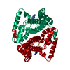
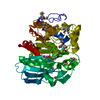
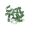
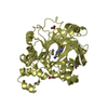
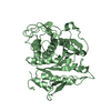
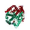
- PDB-5tk6: Structure of the HD-domain phosphohydrolase OxsA with Oxetanocin-... -
+ Open data

- Basic information
| Entry | Database: PDB / ID: 5tk6 | ||||||
|---|---|---|---|---|---|---|---|
| Title | Structure of the HD-domain phosphohydrolase OxsA with Oxetanocin-A diphosphate bound | ||||||
 Components Components | OxsA protein | ||||||
 Keywords Keywords | METAL BINDING PROTEIN / metalloprotein | ||||||
| Function / homology |  Function and homology information Function and homology information4'-phosphooxetanocin A phosphatase / hydrolase activity / metal ion binding Similarity search - Function | ||||||
| Biological species |  Bacillus megaterium (bacteria) Bacillus megaterium (bacteria) | ||||||
| Method |  X-RAY DIFFRACTION / SYNCHROTRON / MOLECULAR REPLACEMENT / Resolution: 1.924 Å X-RAY DIFFRACTION / SYNCHROTRON / MOLECULAR REPLACEMENT / Resolution: 1.924 Å | ||||||
 Authors Authors | Bridwell-Rabb, J. / Drennan, C.L. | ||||||
| Funding support |  United States, 1items United States, 1items
| ||||||
 Citation Citation | Journal: Proc. Natl. Acad. Sci. U.S.A. / Year: 2016 Title: An HD domain phosphohydrolase active site tailored for oxetanocin-A biosynthesis. Authors: Bridwell-Rabb, J. / Kang, G. / Zhong, A. / Liu, H.W. / Drennan, C.L. | ||||||
| History |
|
- Structure visualization
| Structure viewer | Molecule: MolmilJmol/JSmol |
|---|
- Downloads & links
-Download
| PDBx/mmCIF format | 5tk6.cif.gz | 57.1 KB | Display | PDBx/mmCIF format |
|---|---|---|---|---|
| PDB format | pdb5tk6.ent.gz | 39.5 KB | Display | PDB format |
| PDBx/mmJSON format | 5tk6.json.gz | Tree view | PDBx/mmJSON format | |
| Others |  Other downloads Other downloads |
-Validation report
| Arichive directory | https://data.pdbj.org/pub/pdb/validation_reports/tk/5tk6ftp://data.pdbj.org/pub/pdb/validation_reports/tk/5tk6 | HTTPS FTP |
|---|
-Related structure data
| Related structure data | 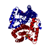 5tk7C  5tk8C  5tk9SC 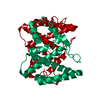 5tkaC C: citing same article ( S: Starting model for refinement |
|---|---|
| Similar structure data |

-Links
 PDBj
PDBj
- Assembly
| Deposited unit | 
| ||||||||
|---|---|---|---|---|---|---|---|---|---|
| 1 | 
| ||||||||
| Unit cell |
|
-Components
| #1: Protein | Mass: 22709.844 Da / Num. of mol.: 1 Source method: isolated from a genetically manipulated source Source: (gene. exp.) Bacillus megaterium (bacteria) / Gene: oxsA / Production host: | ||||
|---|---|---|---|---|---|
| #2: Chemical |   Mass: 24.305 Da / Num. of mol.: 2 / Source method: obtained synthetically / Formula: Mg Mass: 24.305 Da / Num. of mol.: 2 / Source method: obtained synthetically / Formula: Mg#3: Chemical | ChemComp-7D3 / [( | 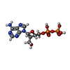  Mass: 411.202 Da / Num. of mol.: 1 / Source method: obtained synthetically / Formula: C10H15N5O9P2 Mass: 411.202 Da / Num. of mol.: 1 / Source method: obtained synthetically / Formula: C10H15N5O9P2#4: Water | ChemComp-HOH / |  Mass: 18.015 Da / Num. of mol.: 76 / Source method: isolated from a natural source / Formula: H2O Mass: 18.015 Da / Num. of mol.: 76 / Source method: isolated from a natural source / Formula: H2O |
-Experimental details
-Experiment
| Experiment | Method: X-RAY DIFFRACTION / Number of used crystals: 1 |
|---|
- Sample preparation
| Crystal | Density Matthews: 2.32 Å3/Da / Density % sol: 46.92 % |
|---|---|
| Crystal grow | Temperature: 298 K / Method: vapor diffusion, hanging drop / pH: 8 Details: 0.1 M TRIS hydrochloride (pH 8.5), 0.2 M magnesium chloride hexahydrate, 30% w/v PEG 400 soaked in a solution of 3 mM OXT-PP in 30% PEG400 for 1 hour Temp details: room temperature |
-Data collection
| Diffraction | Mean temperature: 100 K |
|---|---|
| Diffraction source | Source: SYNCHROTRON / Site: APS / Beamline: 24-ID-C / Wavelength: 0.9792 Å |
| Detector | Type: DECTRIS PILATUS 6M / Detector: PIXEL / Date: Aug 3, 2015 |
| Radiation | Protocol: SINGLE WAVELENGTH / Monochromatic (M) / Laue (L): M / Scattering type: x-ray |
| Radiation wavelength | Wavelength: 0.9792 Å / Relative weight: 1 |
| Reflection | Resolution: 1.92→21.41 Å / Num. obs: 15836 / % possible obs: 99.4 % / Redundancy: 7.3 % / Rmerge(I) obs: 0.068 / Net I/σ(I): 26.2 |
| Reflection shell | Resolution: 1.92→2 Å / Redundancy: 6.9 % / Rmerge(I) obs: 0.903 / Mean I/σ(I) obs: 1.8 / CC1/2: 0.699 / % possible all: 99.7 |
- Processing
| Software |
| |||||||||||||||||||||||||||||||||||||||||||||||||
|---|---|---|---|---|---|---|---|---|---|---|---|---|---|---|---|---|---|---|---|---|---|---|---|---|---|---|---|---|---|---|---|---|---|---|---|---|---|---|---|---|---|---|---|---|---|---|---|---|---|---|
| Refinement | Method to determine structure: MOLECULAR REPLACEMENT Starting model: 5TK9 Resolution: 1.924→21.409 Å / SU ML: 0.23 / Cross valid method: FREE R-VALUE / σ(F): 1.35 / Phase error: 23.57 / Stereochemistry target values: ML
| |||||||||||||||||||||||||||||||||||||||||||||||||
| Solvent computation | Shrinkage radii: 0.9 Å / VDW probe radii: 1.11 Å / Solvent model: FLAT BULK SOLVENT MODEL | |||||||||||||||||||||||||||||||||||||||||||||||||
| Refinement step | Cycle: LAST / Resolution: 1.924→21.409 Å
| |||||||||||||||||||||||||||||||||||||||||||||||||
| Refine LS restraints |
| |||||||||||||||||||||||||||||||||||||||||||||||||
| LS refinement shell |
|
