 Movie
Movie Controller

[English] 日本語
 Yorodumi
- PDB-5tjw: Influenza A virus Nucleoprotein in Complex with Inhibitory Nanobody -
+ Open data

- Basic information
| Entry | Database: PDB / ID: 5tjw | ||||||
|---|---|---|---|---|---|---|---|
| Title | Influenza A virus Nucleoprotein in Complex with Inhibitory Nanobody | ||||||
 Components Components |
| ||||||
 Keywords Keywords | Viral Protein/Immune System / Influenza / nucleoprotein / VHH / inhibitory / Viral Protein-Immune System complex | ||||||
| Function / homology |  Function and homology information Function and homology informationhelical viral capsid / viral penetration into host nucleus / host cell / viral nucleocapsid / ribonucleoprotein complex / symbiont entry into host cell / host cell nucleus / structural molecule activity / RNA binding / identical protein binding Similarity search - Function | ||||||
| Biological species |   Influenza A virus Influenza A virus | ||||||
| Method |  X-RAY DIFFRACTION / SYNCHROTRON / MOLECULAR REPLACEMENT / Resolution: 3.231 Å X-RAY DIFFRACTION / SYNCHROTRON / MOLECULAR REPLACEMENT / Resolution: 3.231 Å | ||||||
 Authors Authors | Hanke, L. / Knockenhauer, K.E. / Ploegh, H.L. / Schwartz, T.U. | ||||||
 Citation Citation | Journal: MBio / Year: 2016 Title: The Antiviral Mechanism of an Influenza A Virus Nucleoprotein-Specific Single-Domain Antibody Fragment. Authors: Hanke, L. / Knockenhauer, K.E. / Brewer, R.C. / van Diest, E. / Schmidt, F.I. / Schwartz, T.U. / Ploegh, H.L. | ||||||
| History |
|
- Structure visualization
| Structure viewer | Molecule: MolmilJmol/JSmol |
|---|
- Downloads & links
-Download
| PDBx/mmCIF format | 5tjw.cif.gz | 127.5 KB | Display | PDBx/mmCIF format |
|---|---|---|---|---|
| PDB format | pdb5tjw.ent.gz | 98.2 KB | Display | PDB format |
| PDBx/mmJSON format | 5tjw.json.gz | Tree view | PDBx/mmJSON format | |
| Others |  Other downloads Other downloads |
-Validation report
| Arichive directory | https://data.pdbj.org/pub/pdb/validation_reports/tj/5tjwftp://data.pdbj.org/pub/pdb/validation_reports/tj/5tjw | HTTPS FTP |
|---|
-Related structure data


-Links
 PDBj
PDBj

- Assembly
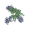
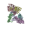
| Deposited unit | 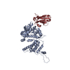
| ||||||||
|---|---|---|---|---|---|---|---|---|---|
| 1 | 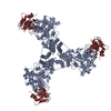
| ||||||||
| Unit cell |
| ||||||||
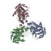
| Details | Hexamer confirmed by gel filtration |
-Components
| #1: Protein | Mass: 57583.012 Da / Num. of mol.: 1 Source method: isolated from a genetically manipulated source Source: (gene. exp.) Influenza A virus (A/WSN/1933(H1N1)) / Strain: A/WSN/1933(H1N1) / Gene: NP / Plasmid: pet30b+ / Production host:  |
|---|---|
| #2: Antibody | Mass: 14934.183 Da / Num. of mol.: 1 Source method: isolated from a genetically manipulated source Source: (gene. exp.) |
| Has protein modification | Y |
-Experimental details
-Experiment
| Experiment | Method: X-RAY DIFFRACTION / Number of used crystals: 1 |
|---|
- Sample preparation
| Crystal | Density Matthews: 2.99 Å3/Da / Density % sol: 58.87 % |
|---|---|
| Crystal grow | Temperature: 291 K / Method: vapor diffusion, hanging drop / pH: 5 Details: 0.1 M sodium acetate, 1.5 M ammonium sulfate, 0.025% (v/v) dichloromethane |
-Data collection
| Diffraction | Mean temperature: 100 K |
|---|---|
| Diffraction source | Source: SYNCHROTRON / Site: APS  / Beamline: 24-ID-C / Wavelength: 0.9778 Å / Beamline: 24-ID-C / Wavelength: 0.9778 Å |
| Detector | Type: DECTRIS PILATUS 6M / Detector: PIXEL / Date: Dec 14, 2014 |
| Radiation | Protocol: SINGLE WAVELENGTH / Monochromatic (M) / Laue (L): M / Scattering type: x-ray |
| Radiation wavelength | Wavelength: 0.9778 Å / Relative weight: 1 |
| Reflection | Resolution: 3.22→97.26 Å / Num. obs: 14189 / % possible obs: 99 % / Redundancy: 9.3 % / CC1/2: 0.996 / Rmerge(I) obs: 0.178 / Net I/σ(I): 15.4 |
| Reflection shell | Resolution: 3.22→3.34 Å / Redundancy: 7.9 % / Mean I/σ(I) obs: 2.1 / CC1/2: 0.687 / % possible all: 100 |
- Processing
| Software |
| |||||||||||||||||||||||||||||||||||||||||||||||||||||||||||||||||||||||||||||
|---|---|---|---|---|---|---|---|---|---|---|---|---|---|---|---|---|---|---|---|---|---|---|---|---|---|---|---|---|---|---|---|---|---|---|---|---|---|---|---|---|---|---|---|---|---|---|---|---|---|---|---|---|---|---|---|---|---|---|---|---|---|---|---|---|---|---|---|---|---|---|---|---|---|---|---|---|---|---|
| Refinement | Method to determine structure: MOLECULAR REPLACEMENT Starting model: 2IQH, 4KRL Resolution: 3.231→97.261 Å / SU ML: 0.38 / Cross valid method: FREE R-VALUE / σ(F): 1.34 / Phase error: 25.47 / Stereochemistry target values: ML
| |||||||||||||||||||||||||||||||||||||||||||||||||||||||||||||||||||||||||||||
| Solvent computation | Shrinkage radii: 0.9 Å / VDW probe radii: 1.11 Å / Solvent model: FLAT BULK SOLVENT MODEL | |||||||||||||||||||||||||||||||||||||||||||||||||||||||||||||||||||||||||||||
| Refinement step | Cycle: LAST / Resolution: 3.231→97.261 Å
| |||||||||||||||||||||||||||||||||||||||||||||||||||||||||||||||||||||||||||||
| Refine LS restraints |
| |||||||||||||||||||||||||||||||||||||||||||||||||||||||||||||||||||||||||||||
| LS refinement shell |
|
