Entry Database : PDB / ID : 5te5Title Crystal structure of Bos taurus opsin regenerated with 6-carbon ring retinal chromophore Rhodopsin Keywords Function / homology Function Domain/homology Component
/ / / / / / / / / / / / / / / / / / / / / / / / / / / / / / / / / / / / / / / / / / / / / / / / / / / / / / / / / / / / / / / / / / / / / / / / Biological species Bos taurus (domestic cattle)Method / / / Resolution : 4.01 Å Authors Gulati, S. / Banerjee, S. / Katayama, K. / Kiser, P.D. / Palczewski, K. Journal : Proc. Natl. Acad. Sci. U.S.A. / Year : 2017Title : Photocyclic behavior of rhodopsin induced by an atypical isomerization mechanism.Authors : Gulati, S. / Jastrzebska, B. / Banerjee, S. / Placeres, A.L. / Miszta, P. / Gao, S. / Gunderson, K. / Tochtrop, G.P. / Filipek, S. / Katayama, K. / Kiser, P.D. / Mogi, M. / Stewart, P.L. / Palczewski, K. History Deposition Sep 20, 2016 Deposition site / Processing site Revision 1.0 Mar 15, 2017 Provider / Type Revision 1.1 Mar 29, 2017 Group Revision 1.2 Apr 5, 2017 Group Revision 1.3 Oct 4, 2023 Group / Database references / Refinement descriptionCategory chem_comp_atom / chem_comp_bond ... chem_comp_atom / chem_comp_bond / database_2 / pdbx_initial_refinement_model Item / _database_2.pdbx_database_accessionRevision 1.4 Nov 20, 2024 Group / Category / pdbx_modification_feature
Show all Show less
 Movie
Movie Controller

 Yorodumi
 Open data
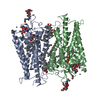
 Basic information
 Components
 Keywords
 Function and homology information
 X-RAY DIFFRACTION /
  Authors
 Citation
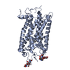
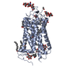
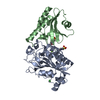
 Structure visualization
 Downloads & links
 Other downloads
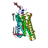


 PDBj
PDBj


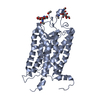
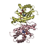
 Assembly

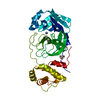
 Mass: 296.446 Da / Num. of mol.: 1 / Source method: obtained synthetically / Formula: C21H28O
 Sample preparation
 / Beamline: 24-ID-C / Wavelength: 0.9792 Å
 Processing