 Movie
Movie Controller


+ Open data

- Basic information
| Entry | Database: PDB / ID: 5smj | |||||||||
|---|---|---|---|---|---|---|---|---|---|---|
| Title | Trypanothione reductase | |||||||||
 Components Components | N(1),N(8)-bis(glutathionyl)spermidine reductase | |||||||||
 Keywords Keywords | OXIDOREDUCTASE / Diamond I04-1 fragment screening / PanDDA / XChemExplorer | |||||||||
| Function / homology |  Function and homology information Function and homology informationtrypanothione-disulfide reductase / trypanothione-disulfide reductase (NADPH) activity / glutathione-disulfide reductase (NADPH) activity / cell redox homeostasis / glutathione metabolic process / flavin adenine dinucleotide binding / cellular response to oxidative stress / mitochondrion / cytosol Similarity search - Function | |||||||||
| Biological species |  | |||||||||
| Method |  X-RAY DIFFRACTION / SYNCHROTRON / FOURIER SYNTHESIS / molecular replacement / Resolution: 1.65 Å X-RAY DIFFRACTION / SYNCHROTRON / FOURIER SYNTHESIS / molecular replacement / Resolution: 1.65 Å | |||||||||
 Authors Authors | Fiorillo, A. / Ilari, A. | |||||||||
| Funding support | European Union,  Italy, 2items Italy, 2items
| |||||||||
 Citation Citation | Journal: Front Mol Biosci / Year: 2022 Title: Innovative Approach for a Classic Target: Fragment Screening on Trypanothione Reductase Reveals New Opportunities for Drug Design. Authors: Fiorillo, A. / Colotti, G. / Exertier, C. / Liuzzi, A. / Seghetti, F. / Salerno, A. / Caciolla, J. / Ilari, A. | |||||||||
| History |
|
- Structure visualization
| Structure viewer | Molecule: MolmilJmol/JSmol |
|---|
- Downloads & links
-Download
| PDBx/mmCIF format | 5smj.cif.gz | 212.7 KB | Display | PDBx/mmCIF format |
|---|---|---|---|---|
| PDB format | pdb5smj.ent.gz | 166.6 KB | Display | PDB format |
| PDBx/mmJSON format | 5smj.json.gz | Tree view | PDBx/mmJSON format | |
| Others |  Other downloads Other downloads |
-Validation report
| Arichive directory | https://data.pdbj.org/pub/pdb/validation_reports/sm/5smjftp://data.pdbj.org/pub/pdb/validation_reports/sm/5smj | HTTPS FTP |
|---|
-Group deposition
| ID | G_1002198 (1 entries) |
|---|---|
| Title | PanDDA analysis group deposition of ground-state model |
| Type | ground state |
| Description | Trypanosoma brucei Trypanothione Reductase screened against the DSiP Fragment Library by X-ray Crystallography at the XChem facility of Diamond Light Source beamline I04-1 |

-Related structure data
| Related structure data |  6rb5S S: Starting model for refinement |
|---|---|
| Similar structure data |
-Links
 PDBj
PDBj


- Assembly
| Deposited unit | 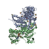
| ||||||||
|---|---|---|---|---|---|---|---|---|---|
| 1 |
| ||||||||
| Unit cell |
|
-Components
-Protein , 1 types, 2 molecules AB
| #1: Protein | Mass: 53497.969 Da / Num. of mol.: 2 Source method: isolated from a genetically manipulated source Source: (gene. exp.)  References: UniProt: A0A3L6KZJ1, trypanothione-disulfide reductase |
|---|
-Non-polymers , 5 types, 547 molecules 


| #2: Chemical |  Mass: 785.550 Da / Num. of mol.: 2 / Source method: obtained synthetically / Formula: C27H33N9O15P2 / Comment: FAD*YM Mass: 785.550 Da / Num. of mol.: 2 / Source method: obtained synthetically / Formula: C27H33N9O15P2 / Comment: FAD*YM#3: Chemical | ChemComp-DMS /  Mass: 78.133 Da / Num. of mol.: 11 / Source method: obtained synthetically / Formula: C2H6OS / Comment: DMSO, precipitant*YM Mass: 78.133 Da / Num. of mol.: 11 / Source method: obtained synthetically / Formula: C2H6OS / Comment: DMSO, precipitant*YM#4: Chemical |  Mass: 24.305 Da / Num. of mol.: 2 / Source method: obtained synthetically / Formula: Mg Mass: 24.305 Da / Num. of mol.: 2 / Source method: obtained synthetically / Formula: Mg#5: Chemical | ChemComp-BR / |  Mass: 79.904 Da / Num. of mol.: 1 / Source method: obtained synthetically / Formula: Br Mass: 79.904 Da / Num. of mol.: 1 / Source method: obtained synthetically / Formula: Br#6: Water | ChemComp-HOH / | Mass: 18.015 Da / Num. of mol.: 531 / Source method: isolated from a natural source / Formula: H2O |
|---|
-Details
| Has protein modification | Y |
|---|
-Experimental details
-Experiment
| Experiment | Method: X-RAY DIFFRACTION / Number of used crystals: 1 |
|---|
- Sample preparation
| Crystal | Density Matthews: 2.3 Å3/Da / Density % sol: 46.41 % |
|---|---|
| Crystal grow | Temperature: 298 K / Method: vapor diffusion, sitting drop / pH: 8 / Details: MPD 22%, PEG 3350 14%, imidazole 40 mM pH 8 |
-Data collection
| Diffraction | Mean temperature: 100 K | |||||||||||||||||||||
|---|---|---|---|---|---|---|---|---|---|---|---|---|---|---|---|---|---|---|---|---|---|---|
| Diffraction source | Source: SYNCHROTRON / Site: Diamond  / Beamline: I04-1 / Wavelength: 0.91264 Å / Beamline: I04-1 / Wavelength: 0.91264 Å | |||||||||||||||||||||
| Detector | Type: DECTRIS PILATUS 6M-F / Detector: PIXEL / Date: Jan 20, 2021 | |||||||||||||||||||||
| Radiation | Protocol: SINGLE WAVELENGTH / Scattering type: x-ray | |||||||||||||||||||||
| Radiation wavelength | Wavelength: 0.91264 Å / Relative weight: 1 | |||||||||||||||||||||
| Reflection | Resolution: 1.65→112.21 Å / Num. obs: 118791 / % possible obs: 98.5 % / CC1/2: 0.99 / Rmerge(I) obs: 0.075 / Net I/σ(I): 15.4 | |||||||||||||||||||||
| Reflection shell |
|
-Phasing
| Phasing | Method: molecular replacement |
|---|
- Processing
| Software |
| |||||||||||||||||||||||||||||||||||||||||||||||||||||||||||||||||||||||||||
|---|---|---|---|---|---|---|---|---|---|---|---|---|---|---|---|---|---|---|---|---|---|---|---|---|---|---|---|---|---|---|---|---|---|---|---|---|---|---|---|---|---|---|---|---|---|---|---|---|---|---|---|---|---|---|---|---|---|---|---|---|---|---|---|---|---|---|---|---|---|---|---|---|---|---|---|---|
| Refinement | Method to determine structure: FOURIER SYNTHESIS Starting model: 6RB5 Resolution: 1.65→78.19 Å / Cor.coef. Fo:Fc: 0.962 / Cor.coef. Fo:Fc free: 0.955 / SU B: 2.799 / SU ML: 0.088 / Cross valid method: THROUGHOUT / σ(F): 0 / ESU R: 0.104 / ESU R Free: 0.099 / Stereochemistry target values: MAXIMUM LIKELIHOOD Details: HYDROGENS HAVE BEEN ADDED IN THE RIDING POSITIONS U VALUES : REFINED INDIVIDUALLY
| |||||||||||||||||||||||||||||||||||||||||||||||||||||||||||||||||||||||||||
| Solvent computation | Ion probe radii: 0.8 Å / Shrinkage radii: 0.8 Å / VDW probe radii: 1.2 Å / Solvent model: MASK | |||||||||||||||||||||||||||||||||||||||||||||||||||||||||||||||||||||||||||
| Displacement parameters | Biso max: 95.3 Å2 / Biso mean: 25.356 Å2 / Biso min: 10.31 Å2
| |||||||||||||||||||||||||||||||||||||||||||||||||||||||||||||||||||||||||||
| Refinement step | Cycle: final / Resolution: 1.65→78.19 Å
| |||||||||||||||||||||||||||||||||||||||||||||||||||||||||||||||||||||||||||
| Refine LS restraints |
| |||||||||||||||||||||||||||||||||||||||||||||||||||||||||||||||||||||||||||
| LS refinement shell | Resolution: 1.65→1.693 Å / Total num. of bins used: 20
|
